Potassium »
PDB 2w0f-2xo0 »
2wj4 »
Potassium in PDB 2wj4: Crystal Structure of the Cofactor-Devoid 1-H-3-Hydroxy-4- Oxoquinaldine 2,4-Dioxygenase (Hod) From Arthrobacter Nitroguajacolicus RU61A Anaerobically Complexed with Its Natural Substrate 1-H-3-Hydroxy-4-Oxoquinaldine
Enzymatic activity of Crystal Structure of the Cofactor-Devoid 1-H-3-Hydroxy-4- Oxoquinaldine 2,4-Dioxygenase (Hod) From Arthrobacter Nitroguajacolicus RU61A Anaerobically Complexed with Its Natural Substrate 1-H-3-Hydroxy-4-Oxoquinaldine
All present enzymatic activity of Crystal Structure of the Cofactor-Devoid 1-H-3-Hydroxy-4- Oxoquinaldine 2,4-Dioxygenase (Hod) From Arthrobacter Nitroguajacolicus RU61A Anaerobically Complexed with Its Natural Substrate 1-H-3-Hydroxy-4-Oxoquinaldine:
1.13.11.48;
1.13.11.48;
Protein crystallography data
The structure of Crystal Structure of the Cofactor-Devoid 1-H-3-Hydroxy-4- Oxoquinaldine 2,4-Dioxygenase (Hod) From Arthrobacter Nitroguajacolicus RU61A Anaerobically Complexed with Its Natural Substrate 1-H-3-Hydroxy-4-Oxoquinaldine, PDB code: 2wj4
was solved by
R.A.Steiner,
with X-Ray Crystallography technique. A brief refinement statistics is given in the table below:
| Resolution Low / High (Å) | 38.84 / 2.10 |
| Space group | P 21 21 21 |
| Cell size a, b, c (Å), α, β, γ (°) | 44.810, 167.200, 167.220, 90.00, 90.00, 90.00 |
| R / Rfree (%) | 17.602 / 20.395 |
Potassium Binding Sites:
The binding sites of Potassium atom in the Crystal Structure of the Cofactor-Devoid 1-H-3-Hydroxy-4- Oxoquinaldine 2,4-Dioxygenase (Hod) From Arthrobacter Nitroguajacolicus RU61A Anaerobically Complexed with Its Natural Substrate 1-H-3-Hydroxy-4-Oxoquinaldine
(pdb code 2wj4). This binding sites where shown within
5.0 Angstroms radius around Potassium atom.
In total 5 binding sites of Potassium where determined in the Crystal Structure of the Cofactor-Devoid 1-H-3-Hydroxy-4- Oxoquinaldine 2,4-Dioxygenase (Hod) From Arthrobacter Nitroguajacolicus RU61A Anaerobically Complexed with Its Natural Substrate 1-H-3-Hydroxy-4-Oxoquinaldine, PDB code: 2wj4:
Jump to Potassium binding site number: 1; 2; 3; 4; 5;
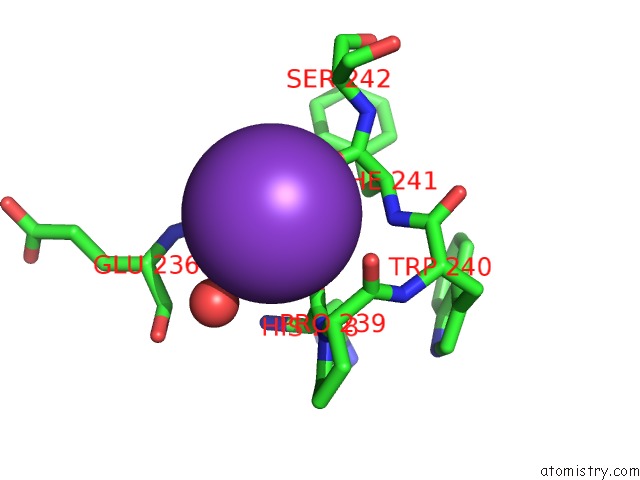
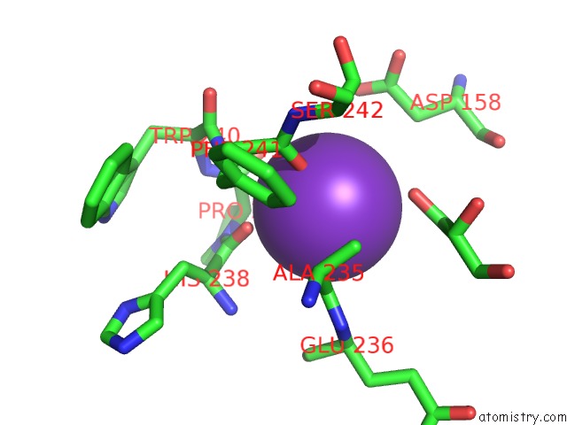
Potassium binding site 1 out of 5 in 2wj4
Go back to
Potassium binding site 1 out
of 5 in the Crystal Structure of the Cofactor-Devoid 1-H-3-Hydroxy-4- Oxoquinaldine 2,4-Dioxygenase (Hod) From Arthrobacter Nitroguajacolicus RU61A Anaerobically Complexed with Its Natural Substrate 1-H-3-Hydroxy-4-Oxoquinaldine
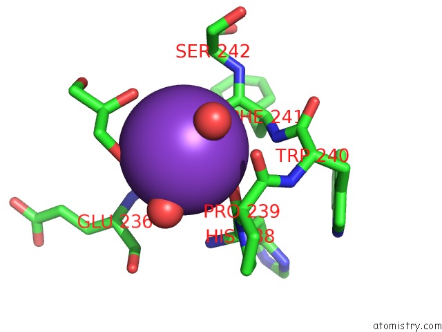
Mono view
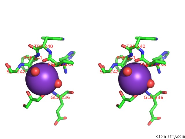
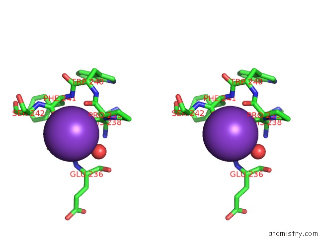
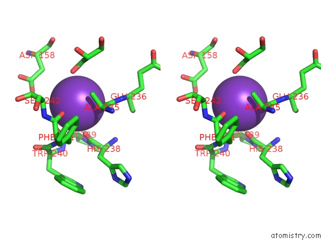
Stereo pair view
Mono view
Stereo pair view
A full contact list of Potassium with other atoms in the K binding
site number 1 of Crystal Structure of the Cofactor-Devoid 1-H-3-Hydroxy-4- Oxoquinaldine 2,4-Dioxygenase (Hod) From Arthrobacter Nitroguajacolicus RU61A Anaerobically Complexed with Its Natural Substrate 1-H-3-Hydroxy-4-Oxoquinaldine within 5.0Å range:
|
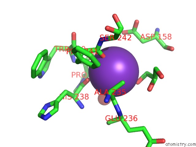
Potassium binding site 2 out of 5 in 2wj4
Go back to
Potassium binding site 2 out
of 5 in the Crystal Structure of the Cofactor-Devoid 1-H-3-Hydroxy-4- Oxoquinaldine 2,4-Dioxygenase (Hod) From Arthrobacter Nitroguajacolicus RU61A Anaerobically Complexed with Its Natural Substrate 1-H-3-Hydroxy-4-Oxoquinaldine
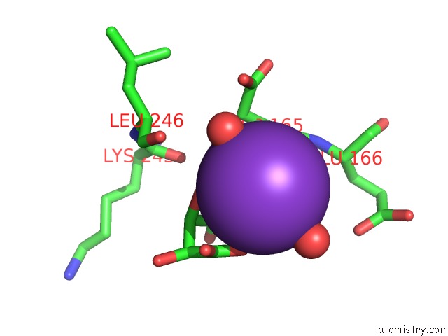
Mono view
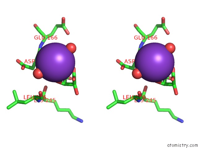
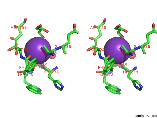
Stereo pair view
Mono view
Stereo pair view
A full contact list of Potassium with other atoms in the K binding
site number 2 of Crystal Structure of the Cofactor-Devoid 1-H-3-Hydroxy-4- Oxoquinaldine 2,4-Dioxygenase (Hod) From Arthrobacter Nitroguajacolicus RU61A Anaerobically Complexed with Its Natural Substrate 1-H-3-Hydroxy-4-Oxoquinaldine within 5.0Å range:
|
Potassium binding site 3 out of 5 in 2wj4
Go back to
Potassium binding site 3 out
of 5 in the Crystal Structure of the Cofactor-Devoid 1-H-3-Hydroxy-4- Oxoquinaldine 2,4-Dioxygenase (Hod) From Arthrobacter Nitroguajacolicus RU61A Anaerobically Complexed with Its Natural Substrate 1-H-3-Hydroxy-4-Oxoquinaldine
Mono view
Stereo pair view
Mono view
Stereo pair view
A full contact list of Potassium with other atoms in the K binding
site number 3 of Crystal Structure of the Cofactor-Devoid 1-H-3-Hydroxy-4- Oxoquinaldine 2,4-Dioxygenase (Hod) From Arthrobacter Nitroguajacolicus RU61A Anaerobically Complexed with Its Natural Substrate 1-H-3-Hydroxy-4-Oxoquinaldine within 5.0Å range:
|
Potassium binding site 4 out of 5 in 2wj4
Go back to
Potassium binding site 4 out
of 5 in the Crystal Structure of the Cofactor-Devoid 1-H-3-Hydroxy-4- Oxoquinaldine 2,4-Dioxygenase (Hod) From Arthrobacter Nitroguajacolicus RU61A Anaerobically Complexed with Its Natural Substrate 1-H-3-Hydroxy-4-Oxoquinaldine
Mono view
Stereo pair view
Mono view
Stereo pair view
A full contact list of Potassium with other atoms in the K binding
site number 4 of Crystal Structure of the Cofactor-Devoid 1-H-3-Hydroxy-4- Oxoquinaldine 2,4-Dioxygenase (Hod) From Arthrobacter Nitroguajacolicus RU61A Anaerobically Complexed with Its Natural Substrate 1-H-3-Hydroxy-4-Oxoquinaldine within 5.0Å range:
|
Potassium binding site 5 out of 5 in 2wj4
Go back to
Potassium binding site 5 out
of 5 in the Crystal Structure of the Cofactor-Devoid 1-H-3-Hydroxy-4- Oxoquinaldine 2,4-Dioxygenase (Hod) From Arthrobacter Nitroguajacolicus RU61A Anaerobically Complexed with Its Natural Substrate 1-H-3-Hydroxy-4-Oxoquinaldine
Mono view
Stereo pair view
Mono view
Stereo pair view
A full contact list of Potassium with other atoms in the K binding
site number 5 of Crystal Structure of the Cofactor-Devoid 1-H-3-Hydroxy-4- Oxoquinaldine 2,4-Dioxygenase (Hod) From Arthrobacter Nitroguajacolicus RU61A Anaerobically Complexed with Its Natural Substrate 1-H-3-Hydroxy-4-Oxoquinaldine within 5.0Å range:
|
Reference:
R.A.Steiner,
H.J.Janssen,
P.Roversi,
A.J.Oakley,
S.Fetzner.
Structural Basis For Cofactor-Independent Dioxygenation of N-Heteroaromatic Compounds at the {Alpha}/{Beta}-Hydrolase Fold. Proc.Natl.Acad.Sci.Usa V. 107 657 2010.
ISSN: ISSN 0027-8424
PubMed: 20080731
DOI: 10.1073/PNAS.0909033107
Page generated: Mon Aug 12 07:29:10 2024
ISSN: ISSN 0027-8424
PubMed: 20080731
DOI: 10.1073/PNAS.0909033107
Last articles
Zn in 9MJ5Zn in 9HNW
Zn in 9G0L
Zn in 9FNE
Zn in 9DZN
Zn in 9E0I
Zn in 9D32
Zn in 9DAK
Zn in 8ZXC
Zn in 8ZUF