Potassium »
PDB 2w0f-2xo0 »
2whv »
Potassium in PDB 2whv: Crystal Structure of Mouse Cadherin-23 EC1-2 (All Cation Binding Sites Occupied By Calcium)
Protein crystallography data
The structure of Crystal Structure of Mouse Cadherin-23 EC1-2 (All Cation Binding Sites Occupied By Calcium), PDB code: 2whv
was solved by
M.Sotomayor,
W.Weihofen,
R.Gaudet,
D.P.Corey,
with X-Ray Crystallography technique. A brief refinement statistics is given in the table below:
| Resolution Low / High (Å) | 22.25 / 2.36 |
| Space group | H 3 2 |
| Cell size a, b, c (Å), α, β, γ (°) | 151.290, 151.290, 136.881, 90.00, 90.00, 120.00 |
| R / Rfree (%) | 20 / 22.1 |
Other elements in 2whv:
The structure of Crystal Structure of Mouse Cadherin-23 EC1-2 (All Cation Binding Sites Occupied By Calcium) also contains other interesting chemical elements:
| Chlorine | (Cl) | 1 atom |
| Calcium | (Ca) | 4 atoms |
Potassium Binding Sites:
The binding sites of Potassium atom in the Crystal Structure of Mouse Cadherin-23 EC1-2 (All Cation Binding Sites Occupied By Calcium)
(pdb code 2whv). This binding sites where shown within
5.0 Angstroms radius around Potassium atom.
In total 4 binding sites of Potassium where determined in the Crystal Structure of Mouse Cadherin-23 EC1-2 (All Cation Binding Sites Occupied By Calcium), PDB code: 2whv:
Jump to Potassium binding site number: 1; 2; 3; 4;
In total 4 binding sites of Potassium where determined in the Crystal Structure of Mouse Cadherin-23 EC1-2 (All Cation Binding Sites Occupied By Calcium), PDB code: 2whv:
Jump to Potassium binding site number: 1; 2; 3; 4;
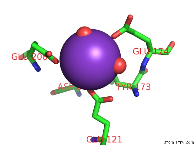
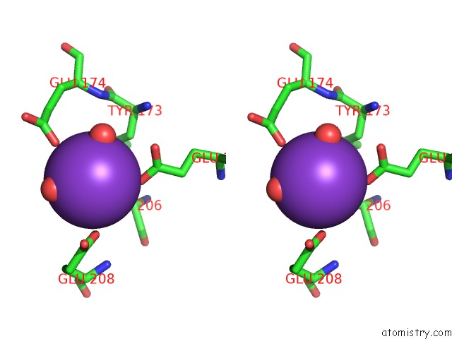
Potassium binding site 1 out of 4 in 2whv
Go back to
Potassium binding site 1 out
of 4 in the Crystal Structure of Mouse Cadherin-23 EC1-2 (All Cation Binding Sites Occupied By Calcium)
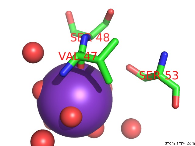
Mono view
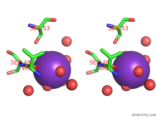
Stereo pair view
Mono view
Stereo pair view
A full contact list of Potassium with other atoms in the K binding
site number 1 of Crystal Structure of Mouse Cadherin-23 EC1-2 (All Cation Binding Sites Occupied By Calcium) within 5.0Å range:
|
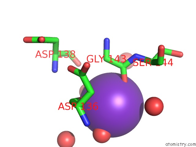
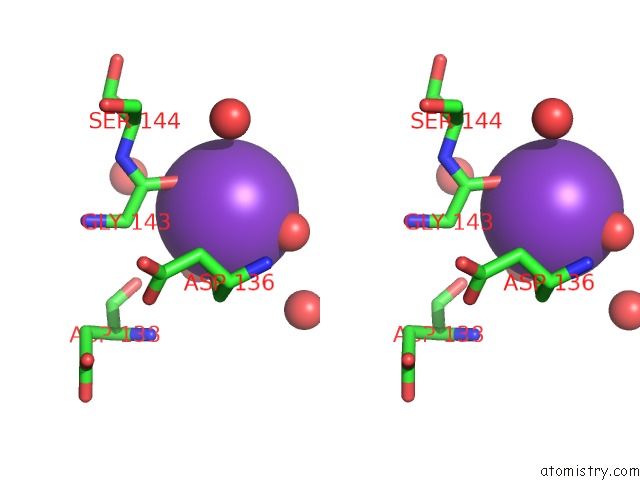
Potassium binding site 2 out of 4 in 2whv
Go back to
Potassium binding site 2 out
of 4 in the Crystal Structure of Mouse Cadherin-23 EC1-2 (All Cation Binding Sites Occupied By Calcium)
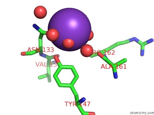
Mono view
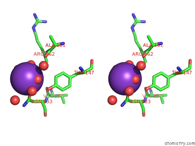
Stereo pair view
Mono view
Stereo pair view
A full contact list of Potassium with other atoms in the K binding
site number 2 of Crystal Structure of Mouse Cadherin-23 EC1-2 (All Cation Binding Sites Occupied By Calcium) within 5.0Å range:
|
Potassium binding site 3 out of 4 in 2whv
Go back to
Potassium binding site 3 out
of 4 in the Crystal Structure of Mouse Cadherin-23 EC1-2 (All Cation Binding Sites Occupied By Calcium)
Mono view
Stereo pair view
Mono view
Stereo pair view
A full contact list of Potassium with other atoms in the K binding
site number 3 of Crystal Structure of Mouse Cadherin-23 EC1-2 (All Cation Binding Sites Occupied By Calcium) within 5.0Å range:
|
Potassium binding site 4 out of 4 in 2whv
Go back to
Potassium binding site 4 out
of 4 in the Crystal Structure of Mouse Cadherin-23 EC1-2 (All Cation Binding Sites Occupied By Calcium)
Mono view
Stereo pair view
Mono view
Stereo pair view
A full contact list of Potassium with other atoms in the K binding
site number 4 of Crystal Structure of Mouse Cadherin-23 EC1-2 (All Cation Binding Sites Occupied By Calcium) within 5.0Å range:
|
Reference:
M.Sotomayor,
W.Weihofen,
R.Gaudet,
D.P.Corey.
Structural Determinants of Cadherin-23 Function in Hearing and Deafness. Neuron V. 66 85 2010.
ISSN: ISSN 0896-6273
PubMed: 20399731
DOI: 10.1016/J.NEURON.2010.03.028
Page generated: Mon Aug 12 07:28:43 2024
ISSN: ISSN 0896-6273
PubMed: 20399731
DOI: 10.1016/J.NEURON.2010.03.028
Last articles
Zn in 9J0NZn in 9J0O
Zn in 9J0P
Zn in 9FJX
Zn in 9EKB
Zn in 9C0F
Zn in 9CAH
Zn in 9CH0
Zn in 9CH3
Zn in 9CH1