Potassium in PDB 2vqv: Structure of HDAC4 Catalytic Domain with A Gain-of-Function Mutation Bound to A Hydroxamic Acid Inhibitor
Protein crystallography data
The structure of Structure of HDAC4 Catalytic Domain with A Gain-of-Function Mutation Bound to A Hydroxamic Acid Inhibitor, PDB code: 2vqv
was solved by
M.J.Bottomley,
P.Lo Surdo,
P.Di Giovine,
A.Cirillo,
R.Scarpelli,
F.Ferrigno,
P.Jones,
P.Neddermann,
R.De Francesco,
C.Steinkuhler,
P.Gallinari,
A.Carfi,
with X-Ray Crystallography technique. A brief refinement statistics is given in the table below:
Resolution Low / High (Å)
|
30.00 /
3.30
|
Space group
|
P 1 21 1
|
Cell size a, b, c (Å), α, β, γ (°)
|
86.524,
70.766,
89.011,
90.00,
108.57,
90.00
|
R / Rfree (%)
|
23.4 /
26.5
|
Other elements in 2vqv:
The structure of Structure of HDAC4 Catalytic Domain with A Gain-of-Function Mutation Bound to A Hydroxamic Acid Inhibitor also contains other interesting chemical elements:
Potassium Binding Sites:
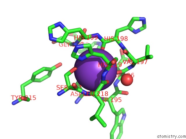
The binding sites of Potassium atom in the Structure of HDAC4 Catalytic Domain with A Gain-of-Function Mutation Bound to A Hydroxamic Acid Inhibitor
(pdb code 2vqv). This binding sites where shown within
5.0 Angstroms radius around Potassium atom.
In total 4 binding sites of Potassium where determined in the
Structure of HDAC4 Catalytic Domain with A Gain-of-Function Mutation Bound to A Hydroxamic Acid Inhibitor, PDB code: 2vqv:
Jump to Potassium binding site number:
1;
2;
3;
4;
Potassium binding site 1 out
of 4 in 2vqv
Go back to
Potassium Binding Sites List in 2vqv
Potassium binding site 1 out
of 4 in the Structure of HDAC4 Catalytic Domain with A Gain-of-Function Mutation Bound to A Hydroxamic Acid Inhibitor
Mono view
Stereo pair view
|
A full contact list of Potassium with other atoms in the K binding
site number 1 of Structure of HDAC4 Catalytic Domain with A Gain-of-Function Mutation Bound to A Hydroxamic Acid Inhibitor within 5.0Å range:
probe
|
atom
|
residue
|
distance (Å)
|
B
|
Occ
|
A:K1407
b:53.6
occ:1.00
|
O
|
A:ASP194
|
2.6
|
30.9
|
1.0
|
O
|
A:HIS198
|
2.6
|
32.4
|
1.0
|
O
|
A:ASP196
|
2.8
|
31.7
|
1.0
|
OD1
|
A:ASP194
|
2.9
|
28.6
|
1.0
|
O
|
A:LEU218
|
3.0
|
33.5
|
1.0
|
OG
|
A:SER217
|
3.1
|
32.2
|
1.0
|
C
|
A:ASP194
|
3.6
|
30.8
|
1.0
|
CG
|
A:ASP194
|
3.7
|
30.1
|
1.0
|
C
|
A:HIS198
|
3.7
|
32.5
|
1.0
|
C
|
A:LEU218
|
3.8
|
33.4
|
1.0
|
C
|
A:ASP196
|
3.8
|
31.7
|
1.0
|
CB
|
A:HIS219
|
3.9
|
34.2
|
1.0
|
N
|
A:ASP196
|
4.0
|
31.4
|
1.0
|
CB
|
A:SER217
|
4.0
|
32.4
|
1.0
|
N
|
A:LEU218
|
4.1
|
32.8
|
1.0
|
CB
|
A:ASP194
|
4.1
|
30.5
|
1.0
|
CA
|
A:SER217
|
4.3
|
32.4
|
1.0
|
CA
|
A:HIS199
|
4.3
|
33.0
|
1.0
|
CA
|
A:ASP196
|
4.3
|
31.6
|
1.0
|
C
|
A:SER217
|
4.3
|
32.6
|
1.0
|
C
|
A:TRP195
|
4.4
|
31.2
|
1.0
|
N
|
A:TRP195
|
4.4
|
30.9
|
1.0
|
N
|
A:HIS219
|
4.4
|
33.8
|
1.0
|
CB
|
A:ASP196
|
4.4
|
31.7
|
1.0
|
N
|
A:GLY200
|
4.4
|
33.1
|
1.0
|
CA
|
A:HIS219
|
4.4
|
34.2
|
1.0
|
ND1
|
A:HIS219
|
4.4
|
34.8
|
1.0
|
CA
|
A:TRP195
|
4.4
|
31.0
|
1.0
|
N
|
A:HIS199
|
4.4
|
32.8
|
1.0
|
CA
|
A:ASP194
|
4.5
|
30.5
|
1.0
|
N
|
A:HIS198
|
4.5
|
32.2
|
1.0
|
O
|
A:HOH2002
|
4.5
|
34.4
|
1.0
|
C
|
A:VAL197
|
4.6
|
32.1
|
1.0
|
CA
|
A:LEU218
|
4.6
|
33.1
|
1.0
|
CG
|
A:HIS219
|
4.6
|
34.5
|
1.0
|
C
|
A:HIS199
|
4.7
|
33.0
|
1.0
|
OD2
|
A:ASP194
|
4.7
|
30.0
|
1.0
|
CE1
|
A:HIS158
|
4.7
|
35.4
|
1.0
|
O
|
A:VAL197
|
4.7
|
32.2
|
1.0
|
CA
|
A:HIS198
|
4.7
|
32.3
|
1.0
|
N
|
A:VAL197
|
4.9
|
31.8
|
1.0
|
OH
|
A:TYR215
|
4.9
|
29.7
|
1.0
|
ND1
|
A:HIS158
|
5.0
|
34.9
|
1.0
|
|
Potassium binding site 2 out
of 4 in 2vqv
Go back to
Potassium Binding Sites List in 2vqv
Potassium binding site 2 out
of 4 in the Structure of HDAC4 Catalytic Domain with A Gain-of-Function Mutation Bound to A Hydroxamic Acid Inhibitor
Mono view
Stereo pair view
|
A full contact list of Potassium with other atoms in the K binding
site number 2 of Structure of HDAC4 Catalytic Domain with A Gain-of-Function Mutation Bound to A Hydroxamic Acid Inhibitor within 5.0Å range:
probe
|
atom
|
residue
|
distance (Å)
|
B
|
Occ
|
A:K1408
b:69.2
occ:1.00
|
O
|
A:PHE207
|
2.7
|
34.9
|
1.0
|
O
|
A:PHE244
|
3.2
|
35.7
|
1.0
|
O
|
A:ASP210
|
3.5
|
36.6
|
1.0
|
O
|
A:VAL213
|
3.6
|
35.8
|
1.0
|
C
|
A:PHE244
|
3.7
|
35.6
|
1.0
|
C
|
A:PHE207
|
3.8
|
34.9
|
1.0
|
CB
|
A:PHE244
|
4.0
|
36.5
|
1.0
|
O
|
A:GLY241
|
4.0
|
39.7
|
1.0
|
N
|
A:ASN245
|
4.1
|
34.7
|
1.0
|
CB
|
A:ASN245
|
4.2
|
33.8
|
1.0
|
CA
|
A:TYR208
|
4.3
|
36.0
|
1.0
|
C
|
A:ASP210
|
4.3
|
36.5
|
1.0
|
CB
|
A:PHE207
|
4.4
|
34.3
|
1.0
|
CA
|
A:PHE244
|
4.4
|
36.4
|
1.0
|
C
|
A:TYR208
|
4.4
|
36.3
|
1.0
|
CA
|
A:ASN245
|
4.5
|
33.9
|
1.0
|
N
|
A:TYR208
|
4.5
|
35.5
|
1.0
|
N
|
A:ASP210
|
4.6
|
36.4
|
1.0
|
C
|
A:VAL213
|
4.7
|
35.6
|
1.0
|
ND2
|
A:ASN245
|
4.7
|
32.9
|
1.0
|
O
|
A:TYR208
|
4.7
|
36.6
|
1.0
|
CA
|
A:PHE207
|
4.7
|
34.5
|
1.0
|
N
|
A:SER209
|
4.8
|
36.4
|
1.0
|
N
|
A:TYR215
|
4.9
|
33.2
|
1.0
|
CA
|
A:GLY241
|
4.9
|
39.8
|
1.0
|
C
|
A:GLY241
|
4.9
|
39.5
|
1.0
|
|
Potassium binding site 3 out
of 4 in 2vqv
Go back to
Potassium Binding Sites List in 2vqv
Potassium binding site 3 out
of 4 in the Structure of HDAC4 Catalytic Domain with A Gain-of-Function Mutation Bound to A Hydroxamic Acid Inhibitor
Mono view
Stereo pair view
|
A full contact list of Potassium with other atoms in the K binding
site number 3 of Structure of HDAC4 Catalytic Domain with A Gain-of-Function Mutation Bound to A Hydroxamic Acid Inhibitor within 5.0Å range:
probe
|
atom
|
residue
|
distance (Å)
|
B
|
Occ
|
B:K1409
b:40.4
occ:1.00
|
O
|
B:HIS198
|
2.5
|
32.5
|
1.0
|
O
|
B:ASP194
|
2.6
|
30.9
|
1.0
|
O
|
B:ASP196
|
2.9
|
31.6
|
1.0
|
OD1
|
B:ASP194
|
2.9
|
28.5
|
1.0
|
OG
|
B:SER217
|
3.0
|
32.1
|
1.0
|
O
|
B:LEU218
|
3.2
|
33.3
|
1.0
|
C
|
B:HIS198
|
3.6
|
32.5
|
1.0
|
C
|
B:ASP194
|
3.7
|
30.7
|
1.0
|
CG
|
B:ASP194
|
3.7
|
30.0
|
1.0
|
C
|
B:ASP196
|
3.8
|
31.6
|
1.0
|
C
|
B:LEU218
|
3.9
|
33.4
|
1.0
|
CB
|
B:HIS219
|
3.9
|
34.3
|
1.0
|
CB
|
B:SER217
|
4.0
|
32.3
|
1.0
|
N
|
B:ASP196
|
4.0
|
31.4
|
1.0
|
CB
|
B:ASP194
|
4.1
|
30.5
|
1.0
|
N
|
B:LEU218
|
4.1
|
32.8
|
1.0
|
CA
|
B:HIS199
|
4.2
|
33.0
|
1.0
|
N
|
B:GLY200
|
4.3
|
33.1
|
1.0
|
CA
|
B:SER217
|
4.3
|
32.3
|
1.0
|
N
|
B:HIS199
|
4.3
|
32.8
|
1.0
|
CA
|
B:ASP196
|
4.3
|
31.7
|
1.0
|
C
|
B:SER217
|
4.4
|
32.5
|
1.0
|
ND1
|
B:HIS219
|
4.4
|
34.8
|
1.0
|
CB
|
B:ASP196
|
4.4
|
31.8
|
1.0
|
CA
|
B:HIS219
|
4.4
|
34.2
|
1.0
|
N
|
B:HIS219
|
4.4
|
33.8
|
1.0
|
C
|
B:TRP195
|
4.5
|
31.2
|
1.0
|
N
|
B:HIS198
|
4.5
|
32.2
|
1.0
|
N
|
B:TRP195
|
4.5
|
30.9
|
1.0
|
CA
|
B:ASP194
|
4.5
|
30.5
|
1.0
|
C
|
B:HIS199
|
4.5
|
32.9
|
1.0
|
CA
|
B:TRP195
|
4.6
|
30.9
|
1.0
|
C
|
B:VAL197
|
4.6
|
32.1
|
1.0
|
CG
|
B:HIS219
|
4.6
|
34.5
|
1.0
|
CE1
|
B:HIS158
|
4.6
|
35.6
|
1.0
|
OD2
|
B:ASP194
|
4.7
|
29.9
|
1.0
|
CA
|
B:LEU218
|
4.7
|
33.1
|
1.0
|
CA
|
B:HIS198
|
4.7
|
32.2
|
1.0
|
O
|
B:VAL197
|
4.7
|
32.2
|
1.0
|
OH
|
B:TYR215
|
4.9
|
29.7
|
1.0
|
N
|
B:VAL197
|
4.9
|
31.7
|
1.0
|
ND1
|
B:HIS158
|
4.9
|
35.0
|
1.0
|
|
Potassium binding site 4 out
of 4 in 2vqv
Go back to
Potassium Binding Sites List in 2vqv
Potassium binding site 4 out
of 4 in the Structure of HDAC4 Catalytic Domain with A Gain-of-Function Mutation Bound to A Hydroxamic Acid Inhibitor
Mono view
Stereo pair view
|
A full contact list of Potassium with other atoms in the K binding
site number 4 of Structure of HDAC4 Catalytic Domain with A Gain-of-Function Mutation Bound to A Hydroxamic Acid Inhibitor within 5.0Å range:
probe
|
atom
|
residue
|
distance (Å)
|
B
|
Occ
|
B:K1410
b:64.4
occ:1.00
|
O
|
B:PHE207
|
2.6
|
34.9
|
1.0
|
O
|
B:PHE244
|
3.4
|
35.5
|
1.0
|
O
|
B:ASP210
|
3.4
|
36.7
|
1.0
|
O
|
B:VAL213
|
3.6
|
35.8
|
1.0
|
C
|
B:PHE207
|
3.7
|
34.9
|
1.0
|
C
|
B:PHE244
|
3.8
|
35.6
|
1.0
|
O
|
B:GLY241
|
4.0
|
39.5
|
1.0
|
CB
|
B:PHE244
|
4.0
|
36.6
|
1.0
|
C
|
B:ASP210
|
4.2
|
36.5
|
1.0
|
N
|
B:ASN245
|
4.2
|
34.7
|
1.0
|
CA
|
B:TYR208
|
4.3
|
36.0
|
1.0
|
CB
|
B:PHE207
|
4.3
|
34.3
|
1.0
|
CB
|
B:ASN245
|
4.3
|
33.8
|
1.0
|
C
|
B:TYR208
|
4.4
|
36.2
|
1.0
|
N
|
B:ASP210
|
4.4
|
36.4
|
1.0
|
N
|
B:TYR208
|
4.5
|
35.5
|
1.0
|
CA
|
B:PHE244
|
4.5
|
36.4
|
1.0
|
CA
|
B:ASN245
|
4.6
|
33.8
|
1.0
|
CA
|
B:PHE207
|
4.7
|
34.5
|
1.0
|
C
|
B:VAL213
|
4.7
|
35.5
|
1.0
|
N
|
B:SER209
|
4.7
|
36.4
|
1.0
|
O
|
B:TYR208
|
4.8
|
36.5
|
1.0
|
CA
|
B:GLY241
|
4.8
|
39.7
|
1.0
|
C
|
B:GLY241
|
4.8
|
39.5
|
1.0
|
ND2
|
B:ASN245
|
4.9
|
32.9
|
1.0
|
CA
|
B:ASP210
|
4.9
|
36.3
|
1.0
|
N
|
B:PRO211
|
5.0
|
36.7
|
1.0
|
N
|
B:TYR215
|
5.0
|
33.2
|
1.0
|
|
Reference:
M.J.Bottomley,
P.Lo Surdo,
P.Di Giovine,
A.Cirillo,
R.Scarpelli,
F.Ferrigno,
P.Jones,
P.Neddermann,
R.De Francesco,
C.Steinkuhler,
P.Gallinari,
A.Carfi.
Structural and Functional Analysis of the Human HDAC4 Catalytic Domain Reveals A Regulatory Structural Zinc-Binding Domain. J.Biol.Chem. V. 283 26694 2008.
ISSN: ISSN 0021-9258
PubMed: 18614528
DOI: 10.1074/JBC.M803514200
Page generated: Mon Aug 12 07:18:48 2024
|