Potassium »
PDB 2qyo-2vxy »
2rdg »
Potassium in PDB 2rdg: Crystal Structure of Staphylococcal Superantigen-Like Protein 11 in Complex with Sialyl Lewis X
Protein crystallography data
The structure of Crystal Structure of Staphylococcal Superantigen-Like Protein 11 in Complex with Sialyl Lewis X, PDB code: 2rdg
was solved by
M.C.Chung,
B.D.Wines,
H.Baker,
R.J.Langley,
E.N.Baker,
J.D.Fraser,
with X-Ray Crystallography technique. A brief refinement statistics is given in the table below:
| Resolution Low / High (Å) | 22.45 / 1.60 |
| Space group | C 1 2 1 |
| Cell size a, b, c (Å), α, β, γ (°) | 97.031, 57.945, 43.462, 90.00, 101.83, 90.00 |
| R / Rfree (%) | 17.4 / 20.7 |
Potassium Binding Sites:
The binding sites of Potassium atom in the Crystal Structure of Staphylococcal Superantigen-Like Protein 11 in Complex with Sialyl Lewis X
(pdb code 2rdg). This binding sites where shown within
5.0 Angstroms radius around Potassium atom.
In total 3 binding sites of Potassium where determined in the Crystal Structure of Staphylococcal Superantigen-Like Protein 11 in Complex with Sialyl Lewis X, PDB code: 2rdg:
Jump to Potassium binding site number: 1; 2; 3;
In total 3 binding sites of Potassium where determined in the Crystal Structure of Staphylococcal Superantigen-Like Protein 11 in Complex with Sialyl Lewis X, PDB code: 2rdg:
Jump to Potassium binding site number: 1; 2; 3;
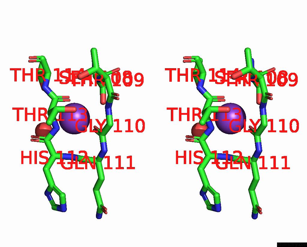
Potassium binding site 1 out of 3 in 2rdg
Go back to
Potassium binding site 1 out
of 3 in the Crystal Structure of Staphylococcal Superantigen-Like Protein 11 in Complex with Sialyl Lewis X
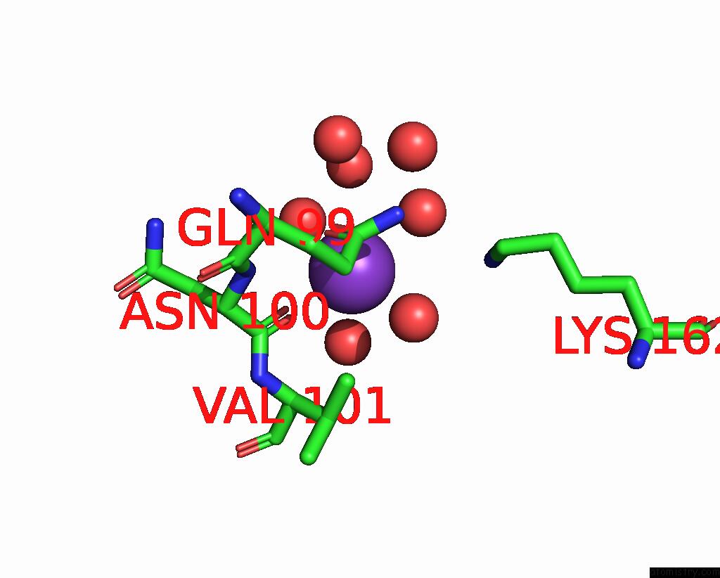
Mono view
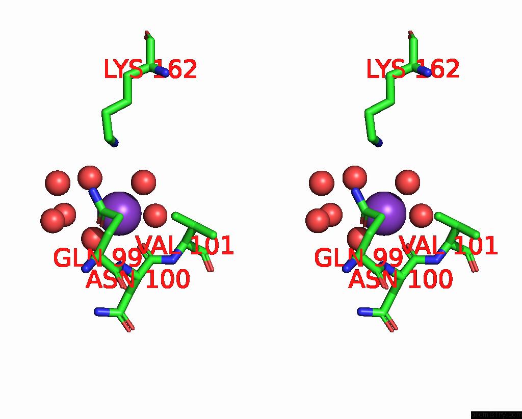
Stereo pair view
Mono view
Stereo pair view
A full contact list of Potassium with other atoms in the K binding
site number 1 of Crystal Structure of Staphylococcal Superantigen-Like Protein 11 in Complex with Sialyl Lewis X within 5.0Å range:
|
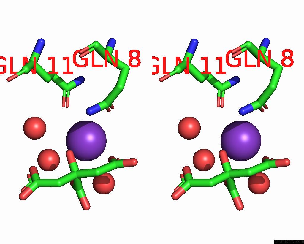
Potassium binding site 2 out of 3 in 2rdg
Go back to
Potassium binding site 2 out
of 3 in the Crystal Structure of Staphylococcal Superantigen-Like Protein 11 in Complex with Sialyl Lewis X

Mono view
Stereo pair view
Mono view
Stereo pair view
A full contact list of Potassium with other atoms in the K binding
site number 2 of Crystal Structure of Staphylococcal Superantigen-Like Protein 11 in Complex with Sialyl Lewis X within 5.0Å range:
|
Potassium binding site 3 out of 3 in 2rdg
Go back to
Potassium binding site 3 out
of 3 in the Crystal Structure of Staphylococcal Superantigen-Like Protein 11 in Complex with Sialyl Lewis X
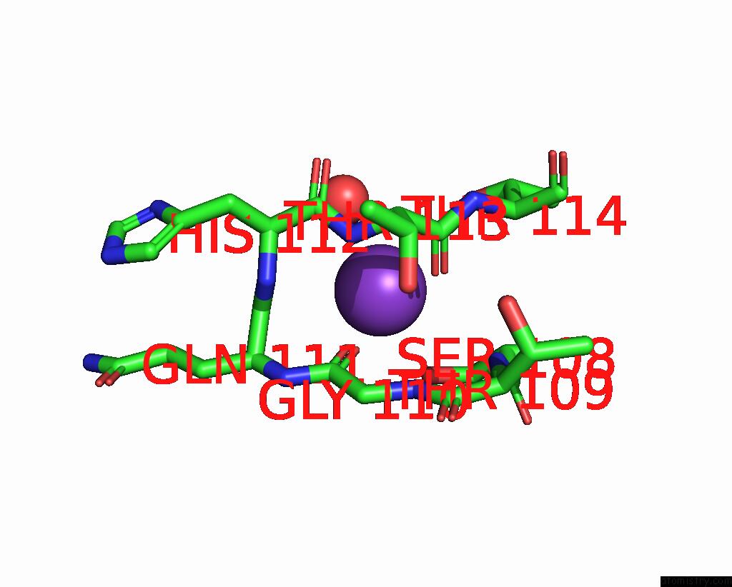
Mono view
Stereo pair view
Mono view
Stereo pair view
A full contact list of Potassium with other atoms in the K binding
site number 3 of Crystal Structure of Staphylococcal Superantigen-Like Protein 11 in Complex with Sialyl Lewis X within 5.0Å range:
|
Reference:
M.C.Chung,
B.D.Wines,
H.Baker,
R.J.Langley,
E.N.Baker,
J.D.Fraser.
The Crystal Structure of Staphylococcal Superantigen-Like Protein 11 in Complex with Sialyl Lewis X Reveals the Mechanism For Cell Binding and Immune Inhibition Mol.Microbiol. V. 66 1342 2007.
ISSN: ISSN 0950-382X
PubMed: 18045383
DOI: 10.1111/J.1365-2958.2007.05989.X
Page generated: Mon Aug 12 06:56:59 2024
ISSN: ISSN 0950-382X
PubMed: 18045383
DOI: 10.1111/J.1365-2958.2007.05989.X
Last articles
Zn in 9J0NZn in 9J0O
Zn in 9J0P
Zn in 9FJX
Zn in 9EKB
Zn in 9C0F
Zn in 9CAH
Zn in 9CH0
Zn in 9CH3
Zn in 9CH1