Potassium »
PDB 2o8l-2qxl »
2qm2 »
Potassium in PDB 2qm2: Putative Hopj Type III Effector Protein From Vibrio Parahaemolyticus
Protein crystallography data
The structure of Putative Hopj Type III Effector Protein From Vibrio Parahaemolyticus, PDB code: 2qm2
was solved by
Y.Kim,
C.Chang,
L.Volkart,
J.Abdullah,
A.Joachimiak,
Midwest Center Forstructural Genomics (Mcsg),
with X-Ray Crystallography technique. A brief refinement statistics is given in the table below:
| Resolution Low / High (Å) | 38.49 / 2.09 |
| Space group | C 2 2 21 |
| Cell size a, b, c (Å), α, β, γ (°) | 87.622, 90.886, 72.445, 90.00, 90.00, 90.00 |
| R / Rfree (%) | 20.9 / 28.1 |
Other elements in 2qm2:
The structure of Putative Hopj Type III Effector Protein From Vibrio Parahaemolyticus also contains other interesting chemical elements:
| Sodium | (Na) | 1 atom |
Potassium Binding Sites:
The binding sites of Potassium atom in the Putative Hopj Type III Effector Protein From Vibrio Parahaemolyticus
(pdb code 2qm2). This binding sites where shown within
5.0 Angstroms radius around Potassium atom.
In total only one binding site of Potassium was determined in the Putative Hopj Type III Effector Protein From Vibrio Parahaemolyticus, PDB code: 2qm2:
In total only one binding site of Potassium was determined in the Putative Hopj Type III Effector Protein From Vibrio Parahaemolyticus, PDB code: 2qm2:
Potassium binding site 1 out of 1 in 2qm2
Go back to
Potassium binding site 1 out
of 1 in the Putative Hopj Type III Effector Protein From Vibrio Parahaemolyticus
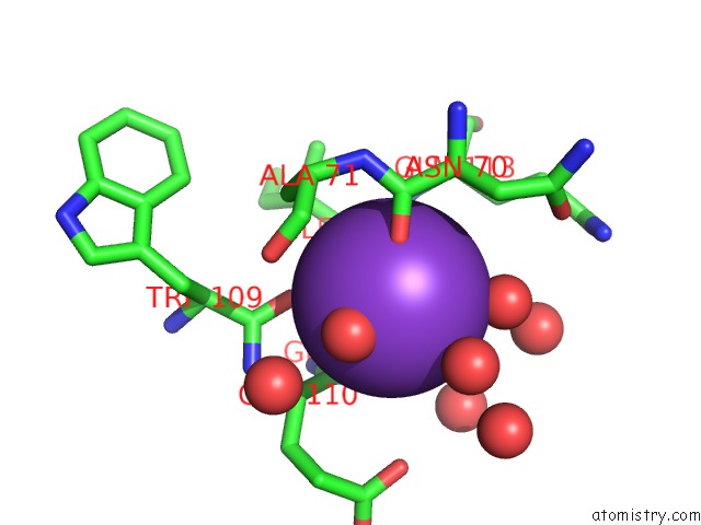
Mono view
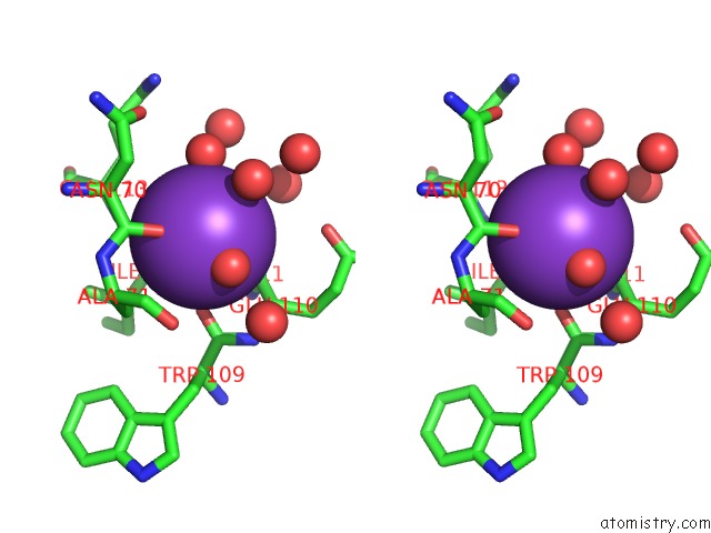
Stereo pair view
Mono view
Stereo pair view
A full contact list of Potassium with other atoms in the K binding
site number 1 of Putative Hopj Type III Effector Protein From Vibrio Parahaemolyticus within 5.0Å range:
|
Reference:
Y.Kim,
C.Chang,
L.Volkart,
J.Abdullah,
A.Joachimiak.
Crystal Structure of Putative Hopj Type III Effector Protein From Vibrio Parahaemolyticus. To Be Published.
Page generated: Sat Aug 9 03:49:45 2025
Last articles
K in 6Q8PK in 6Q8M
K in 6Q7I
K in 6Q8N
K in 6Q0Z
K in 6PZU
K in 6Q01
K in 6Q2C
K in 6PIC
K in 6Q00