Potassium »
PDB 2o8l-2qxl »
2qbz »
Potassium in PDB 2qbz: Structure of the M-Box Riboswitch Aptamer Domain
Protein crystallography data
The structure of Structure of the M-Box Riboswitch Aptamer Domain, PDB code: 2qbz
was solved by
C.E.Dann Iii,
W.C.Winkler,
with X-Ray Crystallography technique. A brief refinement statistics is given in the table below:
| Resolution Low / High (Å) | 47.67 / 2.60 |
| Space group | I 2 2 2 |
| Cell size a, b, c (Å), α, β, γ (°) | 48.655, 101.081, 285.765, 90.00, 90.00, 90.00 |
| R / Rfree (%) | 20.2 / 24.6 |
Other elements in 2qbz:
The structure of Structure of the M-Box Riboswitch Aptamer Domain also contains other interesting chemical elements:
| Magnesium | (Mg) | 6 atoms |
Potassium Binding Sites:
The binding sites of Potassium atom in the Structure of the M-Box Riboswitch Aptamer Domain
(pdb code 2qbz). This binding sites where shown within
5.0 Angstroms radius around Potassium atom.
In total 4 binding sites of Potassium where determined in the Structure of the M-Box Riboswitch Aptamer Domain, PDB code: 2qbz:
Jump to Potassium binding site number: 1; 2; 3; 4;
In total 4 binding sites of Potassium where determined in the Structure of the M-Box Riboswitch Aptamer Domain, PDB code: 2qbz:
Jump to Potassium binding site number: 1; 2; 3; 4;
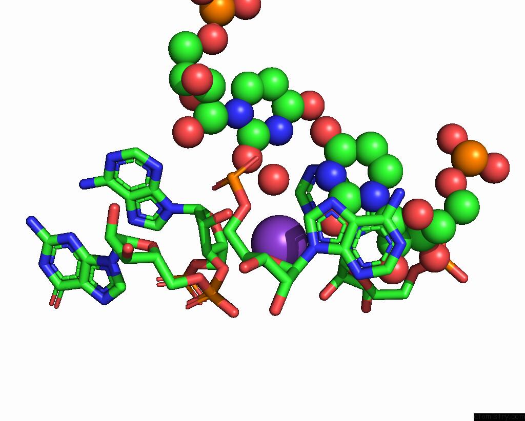
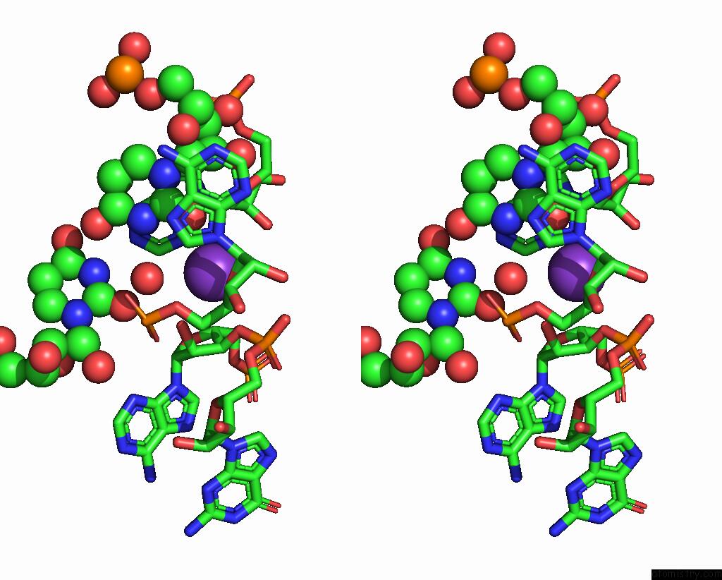
Potassium binding site 1 out of 4 in 2qbz
Go back to
Potassium binding site 1 out
of 4 in the Structure of the M-Box Riboswitch Aptamer Domain
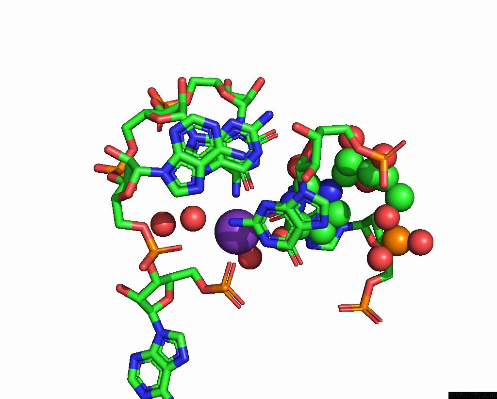
Mono view
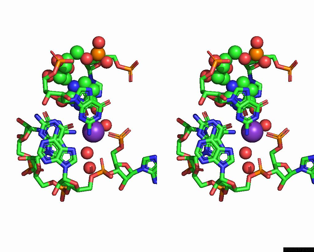
Stereo pair view
Mono view
Stereo pair view
A full contact list of Potassium with other atoms in the K binding
site number 1 of Structure of the M-Box Riboswitch Aptamer Domain within 5.0Å range:
|
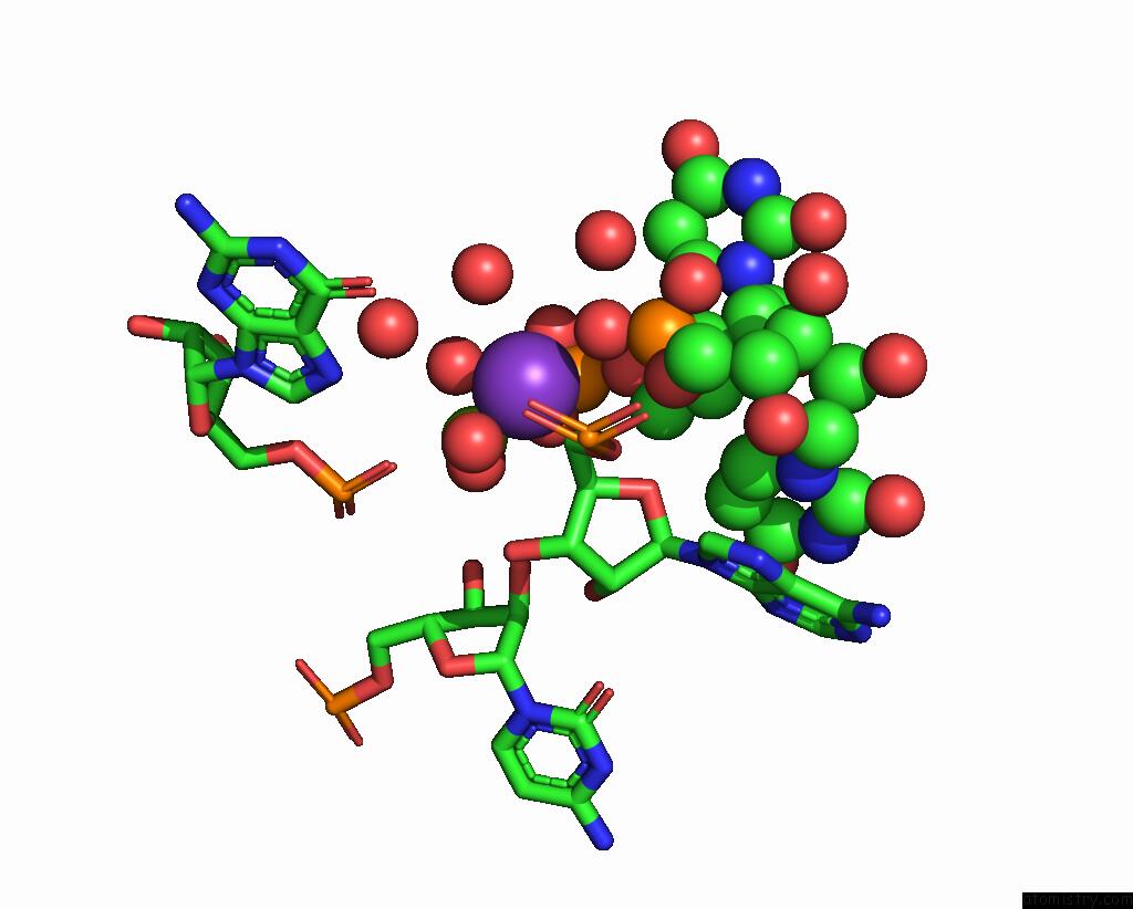
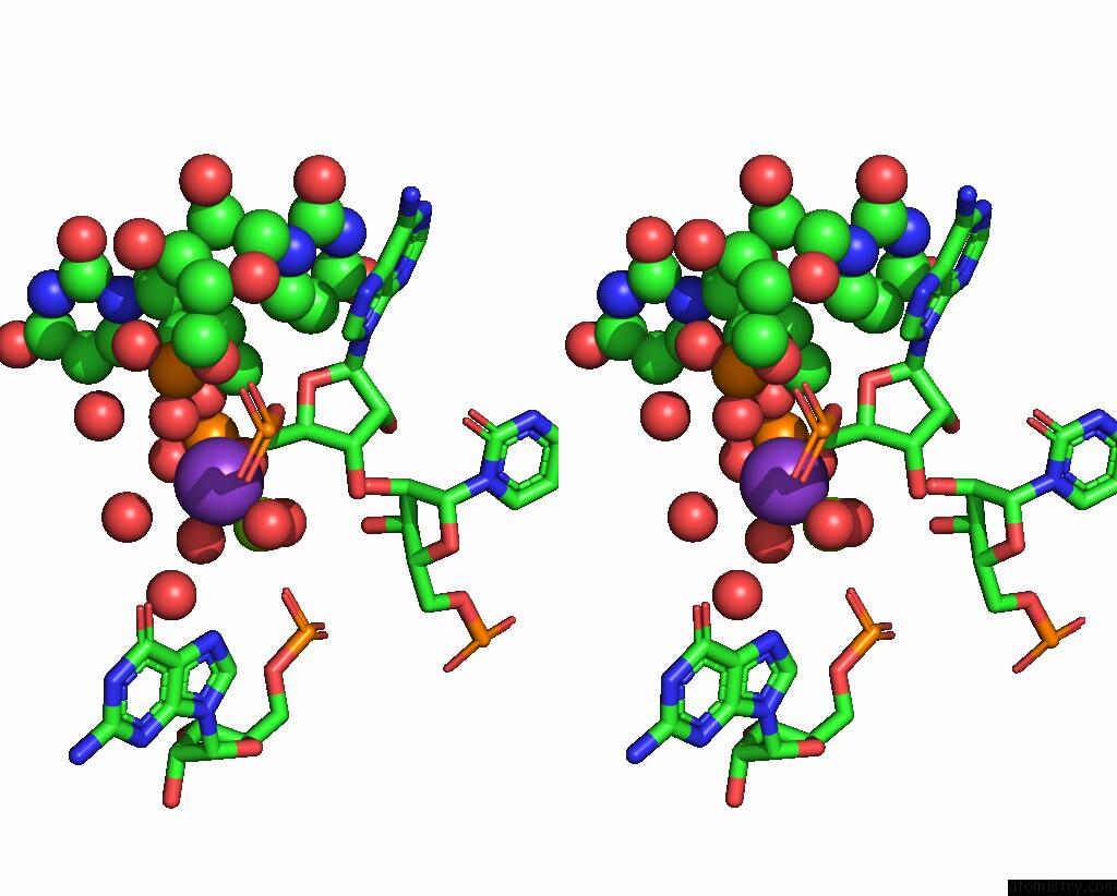
Potassium binding site 2 out of 4 in 2qbz
Go back to
Potassium binding site 2 out
of 4 in the Structure of the M-Box Riboswitch Aptamer Domain
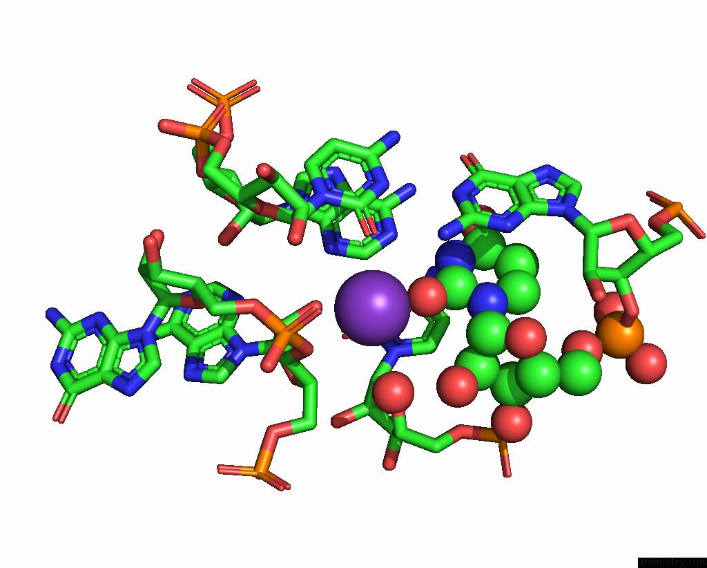
Mono view
Stereo pair view
Mono view
Stereo pair view
A full contact list of Potassium with other atoms in the K binding
site number 2 of Structure of the M-Box Riboswitch Aptamer Domain within 5.0Å range:
|
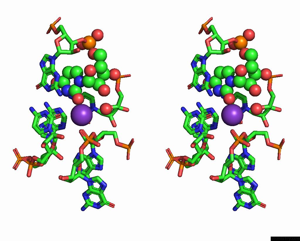
Potassium binding site 3 out of 4 in 2qbz
Go back to
Potassium binding site 3 out
of 4 in the Structure of the M-Box Riboswitch Aptamer Domain
Mono view
Stereo pair view
Mono view
Stereo pair view
A full contact list of Potassium with other atoms in the K binding
site number 3 of Structure of the M-Box Riboswitch Aptamer Domain within 5.0Å range:
|
Potassium binding site 4 out of 4 in 2qbz
Go back to
Potassium binding site 4 out
of 4 in the Structure of the M-Box Riboswitch Aptamer Domain
Mono view
Stereo pair view
Mono view
Stereo pair view
A full contact list of Potassium with other atoms in the K binding
site number 4 of Structure of the M-Box Riboswitch Aptamer Domain within 5.0Å range:
|
Reference:
C.E.Dann Iii,
C.A.Wakeman,
C.L.Sieling,
S.C.Baker,
I.Irnov,
W.C.Winkler.
Structure and Mechanism of A Metal-Sensing Regulatory Rna Cell(Cambridge,Mass.) V. 130 878 2007.
ISSN: ISSN 0092-8674
PubMed: 17803910
DOI: 10.1016/J.CELL.2007.06.051
Page generated: Sat Aug 9 03:48:51 2025
ISSN: ISSN 0092-8674
PubMed: 17803910
DOI: 10.1016/J.CELL.2007.06.051
Last articles
K in 6P45K in 6PC3
K in 6P9V
K in 6P0Y
K in 6OZI
K in 6P1M
K in 6OZJ
K in 6OZR
K in 6OXD
K in 6OF8