Potassium »
PDB 2o8l-2qxl »
2pmu »
Potassium in PDB 2pmu: Crystal Structure of the Dna-Binding Domain of Phop
Protein crystallography data
The structure of Crystal Structure of the Dna-Binding Domain of Phop, PDB code: 2pmu
was solved by
S.Wang,
with X-Ray Crystallography technique. A brief refinement statistics is given in the table below:
| Resolution Low / High (Å) | 20.00 / 1.78 |
| Space group | C 1 2 1 |
| Cell size a, b, c (Å), α, β, γ (°) | 103.884, 101.129, 86.930, 90.00, 126.72, 90.00 |
| R / Rfree (%) | 19.6 / 23.8 |
Other elements in 2pmu:
The structure of Crystal Structure of the Dna-Binding Domain of Phop also contains other interesting chemical elements:
| Chlorine | (Cl) | 4 atoms |
Potassium Binding Sites:
The binding sites of Potassium atom in the Crystal Structure of the Dna-Binding Domain of Phop
(pdb code 2pmu). This binding sites where shown within
5.0 Angstroms radius around Potassium atom.
In total 3 binding sites of Potassium where determined in the Crystal Structure of the Dna-Binding Domain of Phop, PDB code: 2pmu:
Jump to Potassium binding site number: 1; 2; 3;
In total 3 binding sites of Potassium where determined in the Crystal Structure of the Dna-Binding Domain of Phop, PDB code: 2pmu:
Jump to Potassium binding site number: 1; 2; 3;
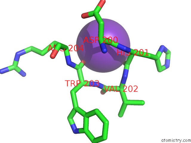
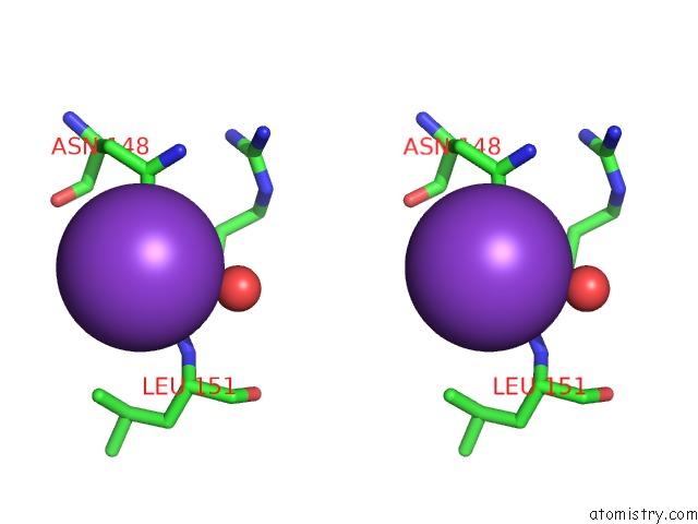
Potassium binding site 1 out of 3 in 2pmu
Go back to
Potassium binding site 1 out
of 3 in the Crystal Structure of the Dna-Binding Domain of Phop

Mono view
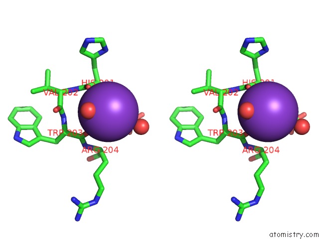
Stereo pair view
Mono view
Stereo pair view
A full contact list of Potassium with other atoms in the K binding
site number 1 of Crystal Structure of the Dna-Binding Domain of Phop within 5.0Å range:
|
Potassium binding site 2 out of 3 in 2pmu
Go back to
Potassium binding site 2 out
of 3 in the Crystal Structure of the Dna-Binding Domain of Phop
Mono view
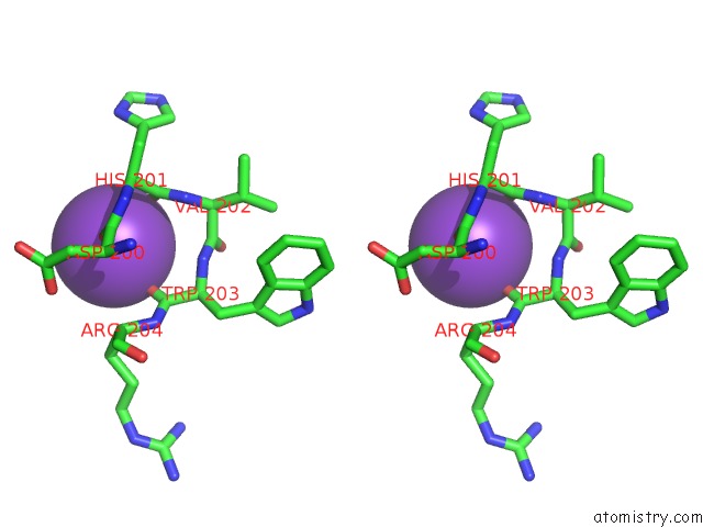
Stereo pair view
Mono view
Stereo pair view
A full contact list of Potassium with other atoms in the K binding
site number 2 of Crystal Structure of the Dna-Binding Domain of Phop within 5.0Å range:
|
Potassium binding site 3 out of 3 in 2pmu
Go back to
Potassium binding site 3 out
of 3 in the Crystal Structure of the Dna-Binding Domain of Phop
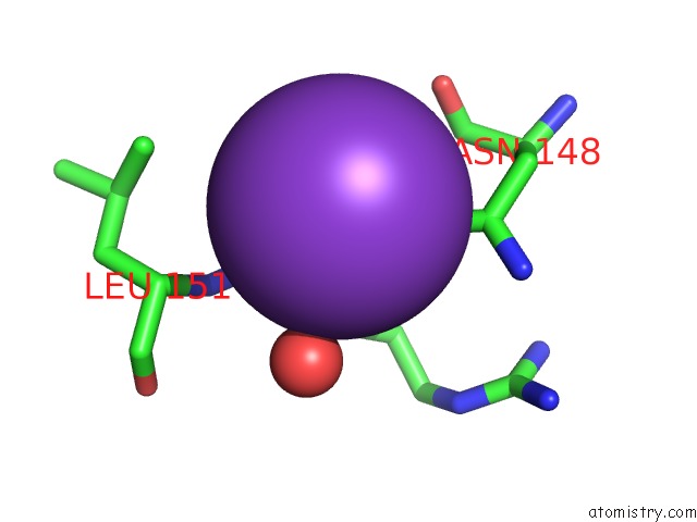
Mono view
Stereo pair view
Mono view
Stereo pair view
A full contact list of Potassium with other atoms in the K binding
site number 3 of Crystal Structure of the Dna-Binding Domain of Phop within 5.0Å range:
|
Reference:
S.Wang,
J.Engohang-Ndong,
I.Smith.
Structure of the Dna-Binding Domain of the Response Regulator Phop From Mycobacterium Tuberculosis Biochemistry V. 46 14751 2007.
ISSN: ISSN 0006-2960
PubMed: 18052041
DOI: 10.1021/BI700970A
Page generated: Mon Aug 12 06:51:18 2024
ISSN: ISSN 0006-2960
PubMed: 18052041
DOI: 10.1021/BI700970A
Last articles
Zn in 9J0NZn in 9J0O
Zn in 9J0P
Zn in 9FJX
Zn in 9EKB
Zn in 9C0F
Zn in 9CAH
Zn in 9CH0
Zn in 9CH3
Zn in 9CH1