Potassium »
PDB 2o8l-2qxl »
2pga »
Potassium in PDB 2pga: X-Ray Structure of the Uridine Phosphorylase From Salmonella Typhimurium in Complex with Inhibitor and Phosphate and Potassium Ion at 1.74 A Resolution
Enzymatic activity of X-Ray Structure of the Uridine Phosphorylase From Salmonella Typhimurium in Complex with Inhibitor and Phosphate and Potassium Ion at 1.74 A Resolution
All present enzymatic activity of X-Ray Structure of the Uridine Phosphorylase From Salmonella Typhimurium in Complex with Inhibitor and Phosphate and Potassium Ion at 1.74 A Resolution:
2.4.2.3;
2.4.2.3;
Protein crystallography data
The structure of X-Ray Structure of the Uridine Phosphorylase From Salmonella Typhimurium in Complex with Inhibitor and Phosphate and Potassium Ion at 1.74 A Resolution, PDB code: 2pga
was solved by
V.I.Timofeev,
A.A.Lashkov,
A.G.Gabdoulkhakov,
C.Betzel,
A.M.Mikhailov,
with X-Ray Crystallography technique. A brief refinement statistics is given in the table below:
| Resolution Low / High (Å) | 6.00 / 1.74 |
| Space group | P 21 21 21 |
| Cell size a, b, c (Å), α, β, γ (°) | 88.790, 124.070, 134.110, 90.00, 90.00, 90.00 |
| R / Rfree (%) | 18.1 / 21 |
Potassium Binding Sites:
The binding sites of Potassium atom in the X-Ray Structure of the Uridine Phosphorylase From Salmonella Typhimurium in Complex with Inhibitor and Phosphate and Potassium Ion at 1.74 A Resolution
(pdb code 2pga). This binding sites where shown within
5.0 Angstroms radius around Potassium atom.
In total 3 binding sites of Potassium where determined in the X-Ray Structure of the Uridine Phosphorylase From Salmonella Typhimurium in Complex with Inhibitor and Phosphate and Potassium Ion at 1.74 A Resolution, PDB code: 2pga:
Jump to Potassium binding site number: 1; 2; 3;
In total 3 binding sites of Potassium where determined in the X-Ray Structure of the Uridine Phosphorylase From Salmonella Typhimurium in Complex with Inhibitor and Phosphate and Potassium Ion at 1.74 A Resolution, PDB code: 2pga:
Jump to Potassium binding site number: 1; 2; 3;
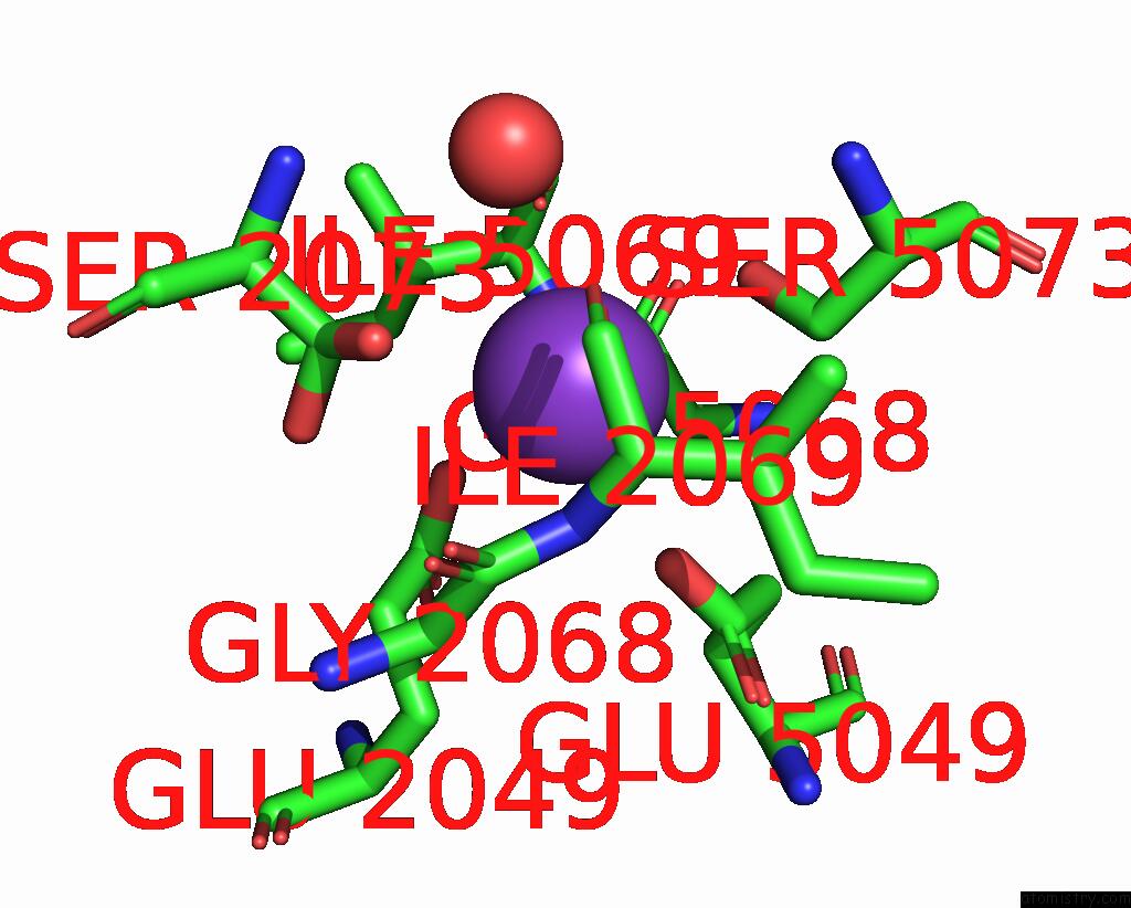
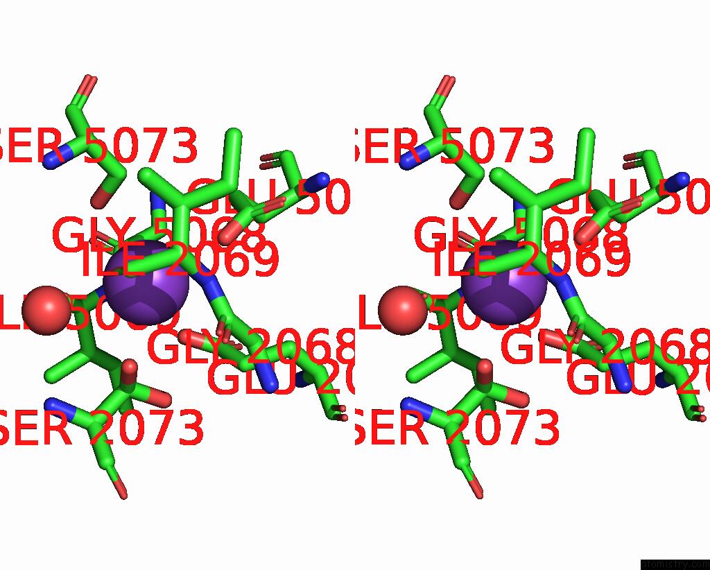
Potassium binding site 1 out of 3 in 2pga
Go back to
Potassium binding site 1 out
of 3 in the X-Ray Structure of the Uridine Phosphorylase From Salmonella Typhimurium in Complex with Inhibitor and Phosphate and Potassium Ion at 1.74 A Resolution
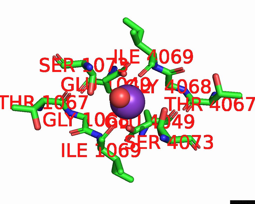
Mono view
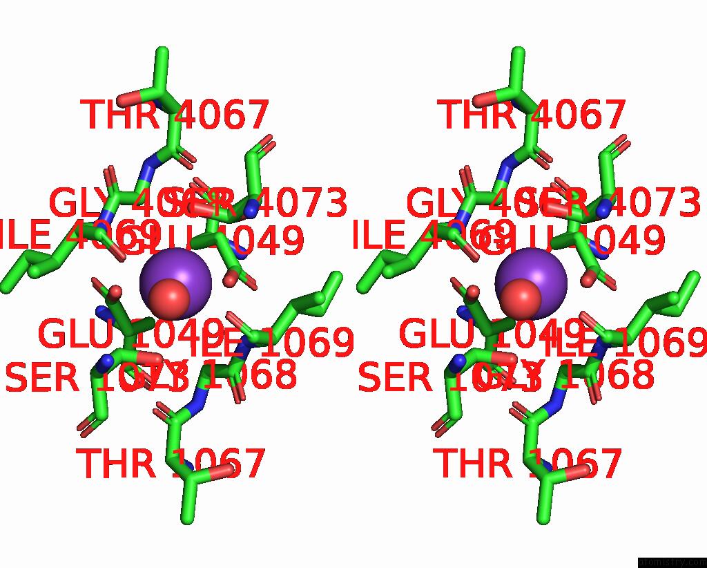
Stereo pair view
Mono view
Stereo pair view
A full contact list of Potassium with other atoms in the K binding
site number 1 of X-Ray Structure of the Uridine Phosphorylase From Salmonella Typhimurium in Complex with Inhibitor and Phosphate and Potassium Ion at 1.74 A Resolution within 5.0Å range:
|
Potassium binding site 2 out of 3 in 2pga
Go back to
Potassium binding site 2 out
of 3 in the X-Ray Structure of the Uridine Phosphorylase From Salmonella Typhimurium in Complex with Inhibitor and Phosphate and Potassium Ion at 1.74 A Resolution
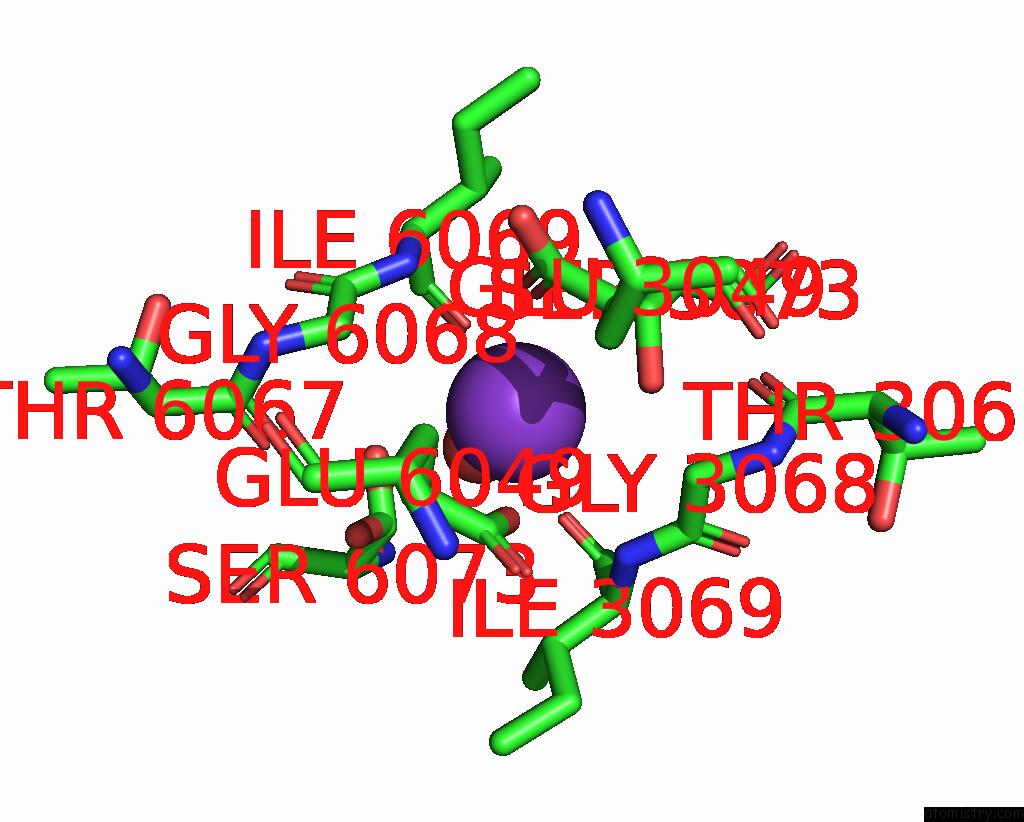
Mono view
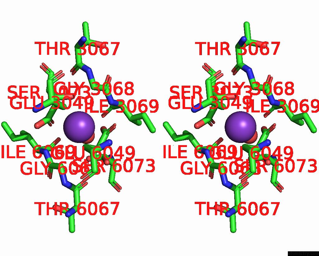
Stereo pair view
Mono view
Stereo pair view
A full contact list of Potassium with other atoms in the K binding
site number 2 of X-Ray Structure of the Uridine Phosphorylase From Salmonella Typhimurium in Complex with Inhibitor and Phosphate and Potassium Ion at 1.74 A Resolution within 5.0Å range:
|
Potassium binding site 3 out of 3 in 2pga
Go back to
Potassium binding site 3 out
of 3 in the X-Ray Structure of the Uridine Phosphorylase From Salmonella Typhimurium in Complex with Inhibitor and Phosphate and Potassium Ion at 1.74 A Resolution
Mono view
Stereo pair view
Mono view
Stereo pair view
A full contact list of Potassium with other atoms in the K binding
site number 3 of X-Ray Structure of the Uridine Phosphorylase From Salmonella Typhimurium in Complex with Inhibitor and Phosphate and Potassium Ion at 1.74 A Resolution within 5.0Å range:
|
Reference:
V.I.Timofeev,
A.A.Lashkov,
A.G.Gabdoulkhakov,
C.Betzel,
A.M.Mikhailov.
X-Ray Structure of the Uridine Phosphorylase From Salmonella Typhimurium in Complex with Inhibitor and Phosphate and Potassium Ion at 1.74 A Resolution To Be Published.
Page generated: Sat Aug 9 03:46:49 2025
Last articles
K in 7OV7K in 7OTJ
K in 7OTB
K in 7OPH
K in 7OUP
K in 7OUE
K in 7OQT
K in 7OQ1
K in 7OT4
K in 7OOU