Potassium »
PDB 2hzv-2o84 »
2nqp »
Potassium in PDB 2nqp: Crystal Structure of Pseudoudirinde Synthase Trua in Complex with Leucyl Trna
Enzymatic activity of Crystal Structure of Pseudoudirinde Synthase Trua in Complex with Leucyl Trna
All present enzymatic activity of Crystal Structure of Pseudoudirinde Synthase Trua in Complex with Leucyl Trna:
5.4.99.12;
5.4.99.12;
Protein crystallography data
The structure of Crystal Structure of Pseudoudirinde Synthase Trua in Complex with Leucyl Trna, PDB code: 2nqp
was solved by
S.Hur,
R.M.Stroud,
with X-Ray Crystallography technique. A brief refinement statistics is given in the table below:
| Resolution Low / High (Å) | 20.00 / 3.50 |
| Space group | P 21 21 21 |
| Cell size a, b, c (Å), α, β, γ (°) | 96.278, 128.585, 159.256, 90.00, 90.00, 90.00 |
| R / Rfree (%) | 24.6 / 27.9 |
Potassium Binding Sites:
The binding sites of Potassium atom in the Crystal Structure of Pseudoudirinde Synthase Trua in Complex with Leucyl Trna
(pdb code 2nqp). This binding sites where shown within
5.0 Angstroms radius around Potassium atom.
In total 6 binding sites of Potassium where determined in the Crystal Structure of Pseudoudirinde Synthase Trua in Complex with Leucyl Trna, PDB code: 2nqp:
Jump to Potassium binding site number: 1; 2; 3; 4; 5; 6;
In total 6 binding sites of Potassium where determined in the Crystal Structure of Pseudoudirinde Synthase Trua in Complex with Leucyl Trna, PDB code: 2nqp:
Jump to Potassium binding site number: 1; 2; 3; 4; 5; 6;
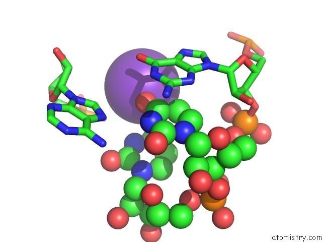
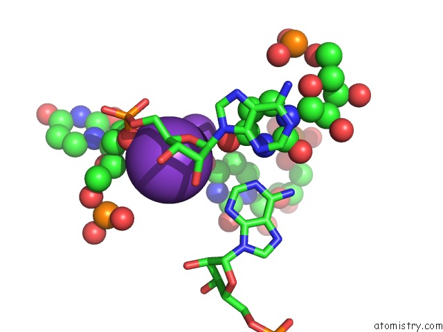
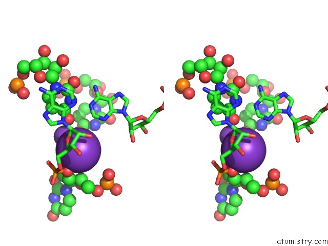
Potassium binding site 1 out of 6 in 2nqp
Go back to
Potassium binding site 1 out
of 6 in the Crystal Structure of Pseudoudirinde Synthase Trua in Complex with Leucyl Trna
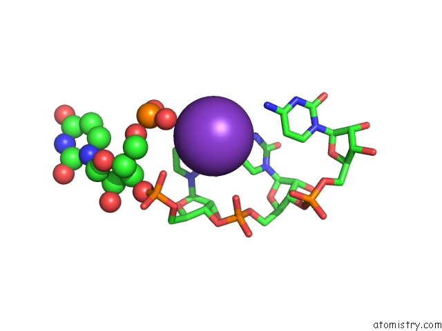
Mono view
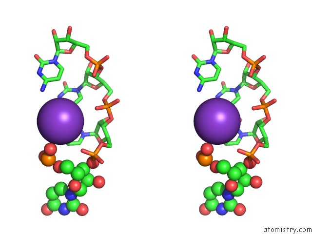
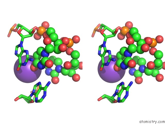
Stereo pair view
Mono view
Stereo pair view
A full contact list of Potassium with other atoms in the K binding
site number 1 of Crystal Structure of Pseudoudirinde Synthase Trua in Complex with Leucyl Trna within 5.0Å range:
|
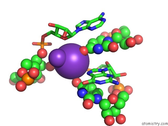
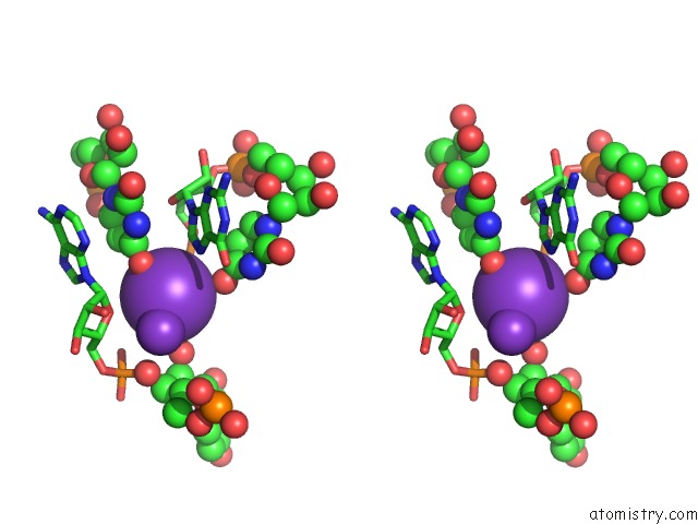
Potassium binding site 2 out of 6 in 2nqp
Go back to
Potassium binding site 2 out
of 6 in the Crystal Structure of Pseudoudirinde Synthase Trua in Complex with Leucyl Trna
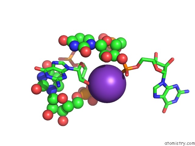
Mono view
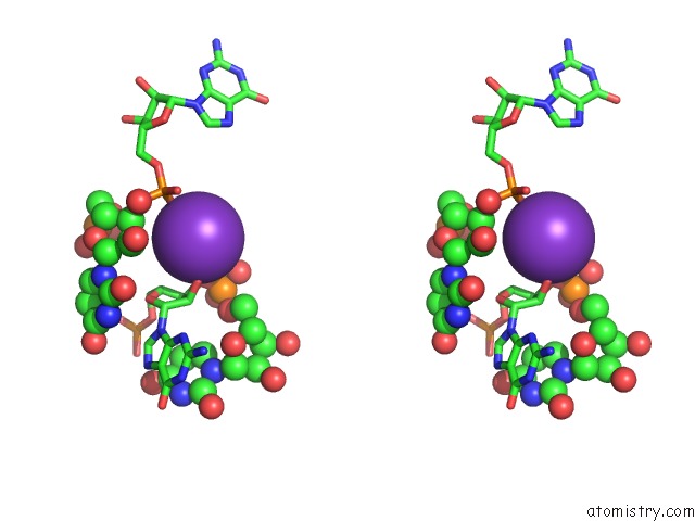
Stereo pair view
Mono view
Stereo pair view
A full contact list of Potassium with other atoms in the K binding
site number 2 of Crystal Structure of Pseudoudirinde Synthase Trua in Complex with Leucyl Trna within 5.0Å range:
|
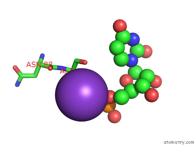
Potassium binding site 3 out of 6 in 2nqp
Go back to
Potassium binding site 3 out
of 6 in the Crystal Structure of Pseudoudirinde Synthase Trua in Complex with Leucyl Trna
Mono view
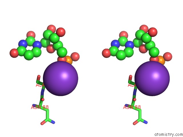
Stereo pair view
Mono view
Stereo pair view
A full contact list of Potassium with other atoms in the K binding
site number 3 of Crystal Structure of Pseudoudirinde Synthase Trua in Complex with Leucyl Trna within 5.0Å range:
|
Potassium binding site 4 out of 6 in 2nqp
Go back to
Potassium binding site 4 out
of 6 in the Crystal Structure of Pseudoudirinde Synthase Trua in Complex with Leucyl Trna
Mono view
Stereo pair view
Mono view
Stereo pair view
A full contact list of Potassium with other atoms in the K binding
site number 4 of Crystal Structure of Pseudoudirinde Synthase Trua in Complex with Leucyl Trna within 5.0Å range:
|
Potassium binding site 5 out of 6 in 2nqp
Go back to
Potassium binding site 5 out
of 6 in the Crystal Structure of Pseudoudirinde Synthase Trua in Complex with Leucyl Trna
Mono view
Stereo pair view
Mono view
Stereo pair view
A full contact list of Potassium with other atoms in the K binding
site number 5 of Crystal Structure of Pseudoudirinde Synthase Trua in Complex with Leucyl Trna within 5.0Å range:
|
Potassium binding site 6 out of 6 in 2nqp
Go back to
Potassium binding site 6 out
of 6 in the Crystal Structure of Pseudoudirinde Synthase Trua in Complex with Leucyl Trna
Mono view
Stereo pair view
Mono view
Stereo pair view
A full contact list of Potassium with other atoms in the K binding
site number 6 of Crystal Structure of Pseudoudirinde Synthase Trua in Complex with Leucyl Trna within 5.0Å range:
|
Reference:
S.Hur,
R.M.Stroud.
How U38, 39, and 40 of Many Trnas Become the Targets For Pseudouridylation By Trua. Mol.Cell V. 26 189 2007.
ISSN: ISSN 1097-2765
PubMed: 17466622
DOI: 10.1016/J.MOLCEL.2007.02.027
Page generated: Mon Aug 12 06:47:58 2024
ISSN: ISSN 1097-2765
PubMed: 17466622
DOI: 10.1016/J.MOLCEL.2007.02.027
Last articles
Zn in 9J0NZn in 9J0O
Zn in 9J0P
Zn in 9FJX
Zn in 9EKB
Zn in 9C0F
Zn in 9CAH
Zn in 9CH0
Zn in 9CH3
Zn in 9CH1