Potassium in PDB 2l8m: Reduced and Co-Bound Cytochrome P450CAM (CYP101A1)
Enzymatic activity of Reduced and Co-Bound Cytochrome P450CAM (CYP101A1)
All present enzymatic activity of Reduced and Co-Bound Cytochrome P450CAM (CYP101A1):
1.14.15.1;
Other elements in 2l8m:
The structure of Reduced and Co-Bound Cytochrome P450CAM (CYP101A1) also contains other interesting chemical elements:
Potassium Binding Sites:
The binding sites of Potassium atom in the Reduced and Co-Bound Cytochrome P450CAM (CYP101A1)
(pdb code 2l8m). This binding sites where shown within
5.0 Angstroms radius around Potassium atom.
In total 4 binding sites of Potassium where determined in the
Reduced and Co-Bound Cytochrome P450CAM (CYP101A1), PDB code: 2l8m:
Jump to Potassium binding site number:
1;
2;
3;
4;
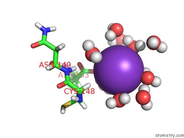
Potassium binding site 1 out
of 4 in 2l8m
Go back to
Potassium Binding Sites List in 2l8m
Potassium binding site 1 out
of 4 in the Reduced and Co-Bound Cytochrome P450CAM (CYP101A1)
Mono view
Stereo pair view
|
A full contact list of Potassium with other atoms in the K binding
site number 1 of Reduced and Co-Bound Cytochrome P450CAM (CYP101A1) within 5.0Å range:
probe
|
atom
|
residue
|
distance (Å)
|
B
|
Occ
|
A:K418
b:0.0
occ:1.00
|
H1
|
A:HOH559
|
2.9
|
0.0
|
1.0
|
O
|
A:HOH843
|
3.0
|
0.0
|
1.0
|
OD1
|
A:ASP153
|
3.1
|
0.0
|
1.0
|
O
|
A:HOH559
|
3.3
|
0.0
|
1.0
|
H1
|
A:HOH843
|
3.4
|
0.0
|
1.0
|
O
|
A:HOH571
|
3.5
|
0.0
|
1.0
|
H2
|
A:HOH843
|
3.5
|
0.0
|
1.0
|
H1
|
A:HOH571
|
3.7
|
0.0
|
1.0
|
H1
|
A:HOH1048
|
3.7
|
0.0
|
1.0
|
H2
|
A:HOH1048
|
3.7
|
0.0
|
1.0
|
H1
|
A:HOH2178
|
3.9
|
0.0
|
1.0
|
H2
|
A:HOH571
|
3.9
|
0.0
|
1.0
|
H2
|
A:HOH471
|
4.0
|
0.0
|
1.0
|
H1
|
A:HOH3247
|
4.1
|
0.0
|
1.0
|
CG
|
A:ASP153
|
4.1
|
0.0
|
1.0
|
O
|
A:HOH1048
|
4.1
|
0.0
|
1.0
|
H2
|
A:HOH559
|
4.2
|
0.0
|
1.0
|
HA
|
A:CYS148
|
4.3
|
0.0
|
1.0
|
OD2
|
A:ASP153
|
4.4
|
0.0
|
1.0
|
O
|
A:HOH2178
|
4.4
|
0.0
|
1.0
|
H2
|
A:HOH3247
|
4.5
|
0.0
|
1.0
|
O
|
A:HOH3247
|
4.6
|
0.0
|
1.0
|
H2
|
A:HOH2178
|
4.7
|
0.0
|
1.0
|
O
|
A:HOH471
|
4.7
|
0.0
|
1.0
|
HB2
|
A:CYS148
|
4.8
|
0.0
|
1.0
|
O
|
A:HOH1496
|
4.9
|
0.0
|
1.0
|
O
|
A:HOH2051
|
4.9
|
0.0
|
1.0
|
H
|
A:ASN149
|
4.9
|
0.0
|
1.0
|
|
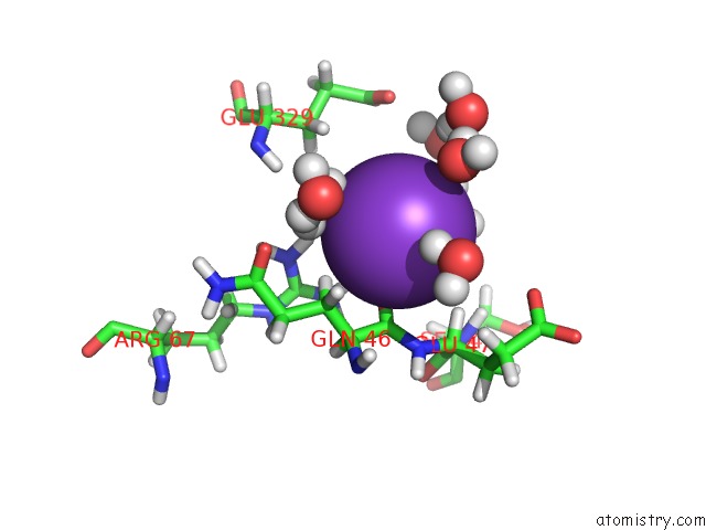
Potassium binding site 2 out
of 4 in 2l8m
Go back to
Potassium Binding Sites List in 2l8m
Potassium binding site 2 out
of 4 in the Reduced and Co-Bound Cytochrome P450CAM (CYP101A1)
Mono view
Stereo pair view
|
A full contact list of Potassium with other atoms in the K binding
site number 2 of Reduced and Co-Bound Cytochrome P450CAM (CYP101A1) within 5.0Å range:
probe
|
atom
|
residue
|
distance (Å)
|
B
|
Occ
|
A:K419
b:0.0
occ:1.00
|
O
|
A:GLN46
|
2.5
|
0.0
|
1.0
|
O
|
A:HOH1905
|
2.8
|
0.0
|
1.0
|
O
|
A:HOH1865
|
2.8
|
0.0
|
1.0
|
H1
|
A:HOH1865
|
3.1
|
0.0
|
1.0
|
HB3
|
A:GLN46
|
3.2
|
0.0
|
1.0
|
HH22
|
A:ARG67
|
3.2
|
0.0
|
1.0
|
H2
|
A:HOH1865
|
3.3
|
0.0
|
1.0
|
C
|
A:GLN46
|
3.4
|
0.0
|
1.0
|
H1
|
A:HOH1905
|
3.4
|
0.0
|
1.0
|
HH12
|
A:ARG67
|
3.5
|
0.0
|
1.0
|
H2
|
A:HOH1905
|
3.5
|
0.0
|
1.0
|
H2
|
A:HOH1886
|
3.6
|
0.0
|
1.0
|
HA
|
A:GLU47
|
3.6
|
0.0
|
1.0
|
HB2
|
A:GLN46
|
3.8
|
0.0
|
1.0
|
CB
|
A:GLN46
|
3.9
|
0.0
|
1.0
|
O
|
A:HOH1912
|
3.9
|
0.0
|
1.0
|
H1
|
A:HOH1912
|
4.1
|
0.0
|
1.0
|
CA
|
A:GLN46
|
4.2
|
0.0
|
1.0
|
N
|
A:GLU47
|
4.2
|
0.0
|
1.0
|
NH2
|
A:ARG67
|
4.2
|
0.0
|
1.0
|
H2
|
A:HOH591
|
4.2
|
0.0
|
1.0
|
O
|
A:HOH1886
|
4.3
|
0.0
|
1.0
|
NH1
|
A:ARG67
|
4.4
|
0.0
|
1.0
|
CA
|
A:GLU47
|
4.5
|
0.0
|
1.0
|
H1
|
A:HOH1886
|
4.6
|
0.0
|
1.0
|
H2
|
A:HOH2409
|
4.7
|
0.0
|
1.0
|
O
|
A:HOH1772
|
4.7
|
0.0
|
1.0
|
HB2
|
A:GLU329
|
4.7
|
0.0
|
1.0
|
HA
|
A:GLN46
|
4.8
|
0.0
|
1.0
|
O
|
A:HOH2409
|
4.8
|
0.0
|
1.0
|
CZ
|
A:ARG67
|
4.8
|
0.0
|
1.0
|
O
|
A:HOH591
|
4.8
|
0.0
|
1.0
|
H2
|
A:HOH1772
|
4.8
|
0.0
|
1.0
|
H2
|
A:HOH1912
|
4.8
|
0.0
|
1.0
|
HH21
|
A:ARG67
|
4.8
|
0.0
|
1.0
|
H
|
A:GLU329
|
5.0
|
0.0
|
1.0
|
H
|
A:SER48
|
5.0
|
0.0
|
1.0
|
|
Potassium binding site 3 out
of 4 in 2l8m
Go back to
Potassium Binding Sites List in 2l8m
Potassium binding site 3 out
of 4 in the Reduced and Co-Bound Cytochrome P450CAM (CYP101A1)
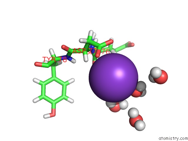
Mono view
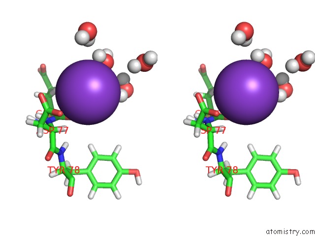
Stereo pair view
|
A full contact list of Potassium with other atoms in the K binding
site number 3 of Reduced and Co-Bound Cytochrome P450CAM (CYP101A1) within 5.0Å range:
probe
|
atom
|
residue
|
distance (Å)
|
B
|
Occ
|
A:K420
b:0.0
occ:1.00
|
O
|
A:GLU76
|
2.7
|
0.0
|
1.0
|
O
|
A:HOH2961
|
3.0
|
0.0
|
1.0
|
H2
|
A:HOH3552
|
3.1
|
0.0
|
1.0
|
H2
|
A:HOH2961
|
3.2
|
0.0
|
1.0
|
HB3
|
A:GLU76
|
3.4
|
0.0
|
1.0
|
C
|
A:GLU76
|
3.5
|
0.0
|
1.0
|
H1
|
A:HOH3570
|
3.5
|
0.0
|
1.0
|
H1
|
A:HOH3552
|
3.7
|
0.0
|
1.0
|
O
|
A:HOH3570
|
3.7
|
0.0
|
1.0
|
O
|
A:HOH3552
|
3.7
|
0.0
|
1.0
|
H1
|
A:HOH2961
|
3.8
|
0.0
|
1.0
|
HA
|
A:ASP77
|
3.8
|
0.0
|
1.0
|
H2
|
A:HOH3570
|
4.0
|
0.0
|
1.0
|
N
|
A:ASP77
|
4.1
|
0.0
|
1.0
|
H1
|
A:HOH3460
|
4.2
|
0.0
|
1.0
|
CB
|
A:GLU76
|
4.4
|
0.0
|
1.0
|
CA
|
A:GLU76
|
4.4
|
0.0
|
1.0
|
CA
|
A:ASP77
|
4.4
|
0.0
|
1.0
|
H1
|
A:HOH3637
|
4.5
|
0.0
|
1.0
|
CG
|
A:ASP77
|
4.6
|
0.0
|
1.0
|
OD1
|
A:ASP77
|
4.6
|
0.0
|
1.0
|
HA
|
A:GLU76
|
4.6
|
0.0
|
1.0
|
OD2
|
A:ASP77
|
4.6
|
0.0
|
1.0
|
H
|
A:ASP77
|
4.7
|
0.0
|
1.0
|
H2
|
A:HOH2481
|
4.8
|
0.0
|
1.0
|
HG2
|
A:GLU76
|
4.9
|
0.0
|
1.0
|
O
|
A:HOH3460
|
4.9
|
0.0
|
1.0
|
H
|
A:TYR78
|
4.9
|
0.0
|
1.0
|
|
Potassium binding site 4 out
of 4 in 2l8m
Go back to
Potassium Binding Sites List in 2l8m
Potassium binding site 4 out
of 4 in the Reduced and Co-Bound Cytochrome P450CAM (CYP101A1)
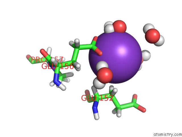
Mono view
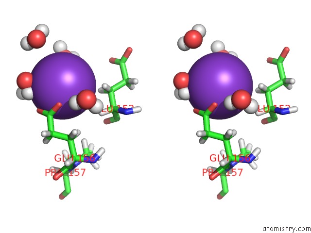
Stereo pair view
|
A full contact list of Potassium with other atoms in the K binding
site number 4 of Reduced and Co-Bound Cytochrome P450CAM (CYP101A1) within 5.0Å range:
probe
|
atom
|
residue
|
distance (Å)
|
B
|
Occ
|
A:K421
b:0.0
occ:1.00
|
OE1
|
A:GLU156
|
2.7
|
0.0
|
1.0
|
OE2
|
A:GLU156
|
2.8
|
0.0
|
1.0
|
H2
|
A:HOH1044
|
2.9
|
0.0
|
1.0
|
O
|
A:HOH1012
|
2.9
|
0.0
|
1.0
|
CD
|
A:GLU156
|
3.0
|
0.0
|
1.0
|
O
|
A:HOH1044
|
3.0
|
0.0
|
1.0
|
O
|
A:HOH1811
|
3.1
|
0.0
|
1.0
|
H1
|
A:HOH1044
|
3.1
|
0.0
|
1.0
|
H1
|
A:HOH1811
|
3.1
|
0.0
|
1.0
|
HB2
|
A:GLU152
|
3.3
|
0.0
|
1.0
|
H2
|
A:HOH1012
|
3.4
|
0.0
|
1.0
|
HB3
|
A:GLU152
|
3.5
|
0.0
|
1.0
|
H1
|
A:HOH1012
|
3.7
|
0.0
|
1.0
|
H1
|
A:HOH1702
|
3.8
|
0.0
|
1.0
|
HA
|
A:GLU152
|
3.9
|
0.0
|
1.0
|
CB
|
A:GLU152
|
4.0
|
0.0
|
1.0
|
H2
|
A:HOH1811
|
4.0
|
0.0
|
1.0
|
HB3
|
A:GLU156
|
4.2
|
0.0
|
1.0
|
O
|
A:HOH2142
|
4.3
|
0.0
|
1.0
|
CG
|
A:GLU156
|
4.4
|
0.0
|
1.0
|
H2
|
A:HOH2142
|
4.4
|
0.0
|
1.0
|
O
|
A:HOH1702
|
4.5
|
0.0
|
1.0
|
CA
|
A:GLU152
|
4.6
|
0.0
|
1.0
|
H2
|
A:HOH1702
|
4.7
|
0.0
|
1.0
|
H1
|
A:HOH2142
|
4.7
|
0.0
|
1.0
|
HG2
|
A:GLU156
|
4.8
|
0.0
|
1.0
|
CB
|
A:GLU156
|
4.8
|
0.0
|
1.0
|
HD3
|
A:PRO157
|
4.9
|
0.0
|
1.0
|
OE1
|
A:GLU152
|
5.0
|
0.0
|
1.0
|
|
Reference:
E.K.Asciutto,
M.Dang,
S.S.Pochapsky,
J.D.Madura,
T.C.Pochapsky.
Experimentally Restrained Molecular Dynamics Simulations For Characterizing the Open States of Cytochrome P450(Cam). Biochemistry V. 50 1664 2011.
ISSN: ISSN 0006-2960
PubMed: 21265500
DOI: 10.1021/BI101820D
Page generated: Mon Aug 12 06:46:44 2024
|