Potassium »
PDB 2hzv-2o84 »
2j41 »
Potassium in PDB 2j41: Crystal Structure of Staphylococcus Aureus Guanylate Monophosphate Kinase
Enzymatic activity of Crystal Structure of Staphylococcus Aureus Guanylate Monophosphate Kinase
All present enzymatic activity of Crystal Structure of Staphylococcus Aureus Guanylate Monophosphate Kinase:
2.7.4.8;
2.7.4.8;
Protein crystallography data
The structure of Crystal Structure of Staphylococcus Aureus Guanylate Monophosphate Kinase, PDB code: 2j41
was solved by
K.El Omari,
B.Dhaliwal,
M.Lockyer,
I.Charles,
A.R.Hawkins,
D.K.Stammers,
with X-Ray Crystallography technique. A brief refinement statistics is given in the table below:
| Resolution Low / High (Å) | 29.40 / 1.9 |
| Space group | P 1 21 1 |
| Cell size a, b, c (Å), α, β, γ (°) | 70.012, 93.986, 83.936, 90.00, 110.59, 90.00 |
| R / Rfree (%) | 20.7 / 23.9 |
Potassium Binding Sites:
The binding sites of Potassium atom in the Crystal Structure of Staphylococcus Aureus Guanylate Monophosphate Kinase
(pdb code 2j41). This binding sites where shown within
5.0 Angstroms radius around Potassium atom.
In total 3 binding sites of Potassium where determined in the Crystal Structure of Staphylococcus Aureus Guanylate Monophosphate Kinase, PDB code: 2j41:
Jump to Potassium binding site number: 1; 2; 3;
In total 3 binding sites of Potassium where determined in the Crystal Structure of Staphylococcus Aureus Guanylate Monophosphate Kinase, PDB code: 2j41:
Jump to Potassium binding site number: 1; 2; 3;
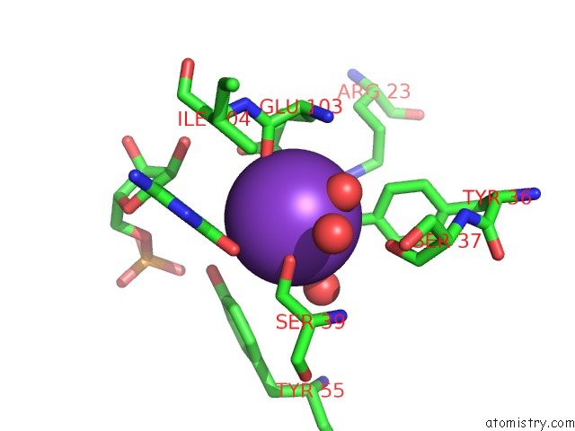
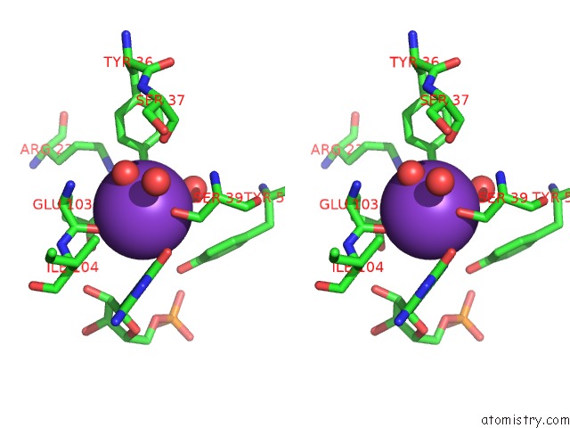
Potassium binding site 1 out of 3 in 2j41
Go back to
Potassium binding site 1 out
of 3 in the Crystal Structure of Staphylococcus Aureus Guanylate Monophosphate Kinase
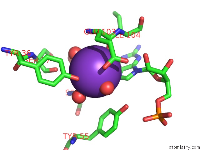
Mono view
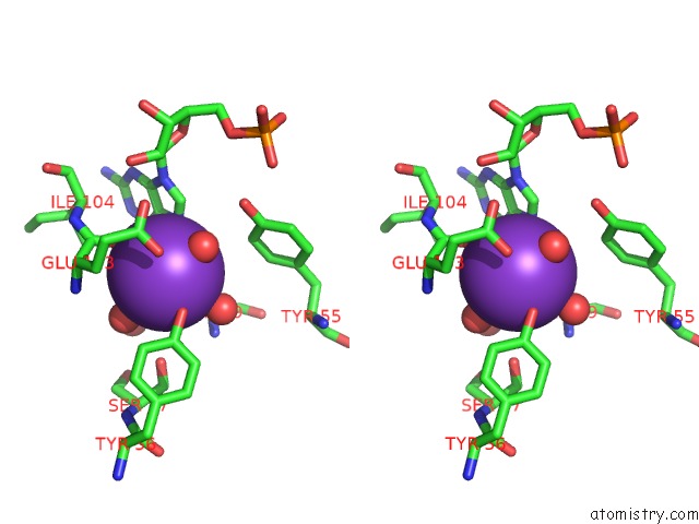
Stereo pair view
Mono view
Stereo pair view
A full contact list of Potassium with other atoms in the K binding
site number 1 of Crystal Structure of Staphylococcus Aureus Guanylate Monophosphate Kinase within 5.0Å range:
|
Potassium binding site 2 out of 3 in 2j41
Go back to
Potassium binding site 2 out
of 3 in the Crystal Structure of Staphylococcus Aureus Guanylate Monophosphate Kinase
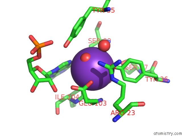
Mono view
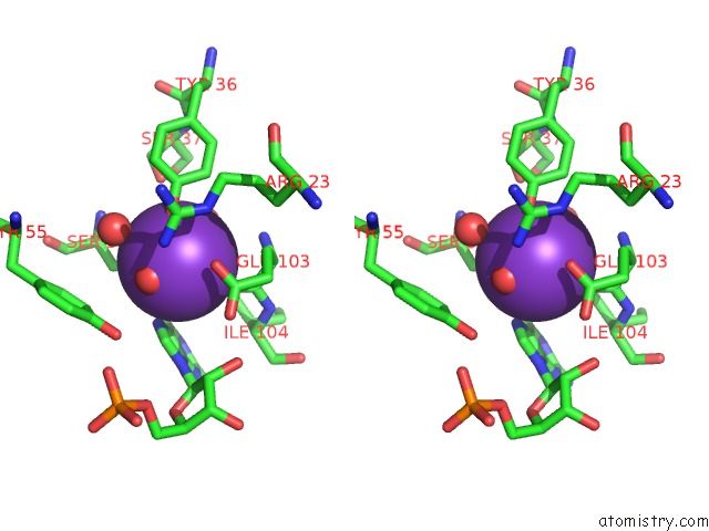
Stereo pair view
Mono view
Stereo pair view
A full contact list of Potassium with other atoms in the K binding
site number 2 of Crystal Structure of Staphylococcus Aureus Guanylate Monophosphate Kinase within 5.0Å range:
|
Potassium binding site 3 out of 3 in 2j41
Go back to
Potassium binding site 3 out
of 3 in the Crystal Structure of Staphylococcus Aureus Guanylate Monophosphate Kinase
Mono view
Stereo pair view
Mono view
Stereo pair view
A full contact list of Potassium with other atoms in the K binding
site number 3 of Crystal Structure of Staphylococcus Aureus Guanylate Monophosphate Kinase within 5.0Å range:
|
Reference:
K.El Omari,
B.Dhaliwal,
M.Lockyer,
I.Charles,
A.R.Hawkins,
D.K.Stammers.
Structure of Staphylococcus Aureus Guanylate Monophosphate Kinase Acta Crystallogr.,Sect.F V. 62 949 2006.
ISSN: ISSN 1744-3091
PubMed: 17012781
DOI: 10.1107/S174430910603613X
Page generated: Mon Aug 12 06:44:08 2024
ISSN: ISSN 1744-3091
PubMed: 17012781
DOI: 10.1107/S174430910603613X
Last articles
Zn in 9J0NZn in 9J0O
Zn in 9J0P
Zn in 9FJX
Zn in 9EKB
Zn in 9C0F
Zn in 9CAH
Zn in 9CH0
Zn in 9CH3
Zn in 9CH1