Potassium »
PDB 2fxi-2hw8 »
2gz5 »
Potassium in PDB 2gz5: Human Type 1 Methionine Aminopeptidase in Complex with Ovalicin at 1.1 Ang
Enzymatic activity of Human Type 1 Methionine Aminopeptidase in Complex with Ovalicin at 1.1 Ang
All present enzymatic activity of Human Type 1 Methionine Aminopeptidase in Complex with Ovalicin at 1.1 Ang:
3.4.11.18;
3.4.11.18;
Protein crystallography data
The structure of Human Type 1 Methionine Aminopeptidase in Complex with Ovalicin at 1.1 Ang, PDB code: 2gz5
was solved by
A.Addlagatta,
B.W.Matthews,
with X-Ray Crystallography technique. A brief refinement statistics is given in the table below:
| Resolution Low / High (Å) | 20.00 / 1.10 |
| Space group | P 1 21 1 |
| Cell size a, b, c (Å), α, β, γ (°) | 47.568, 77.689, 48.644, 90.00, 90.63, 90.00 |
| R / Rfree (%) | 13.5 / 14.5 |
Other elements in 2gz5:
The structure of Human Type 1 Methionine Aminopeptidase in Complex with Ovalicin at 1.1 Ang also contains other interesting chemical elements:
| Cobalt | (Co) | 3 atoms |
Potassium Binding Sites:
The binding sites of Potassium atom in the Human Type 1 Methionine Aminopeptidase in Complex with Ovalicin at 1.1 Ang
(pdb code 2gz5). This binding sites where shown within
5.0 Angstroms radius around Potassium atom.
In total only one binding site of Potassium was determined in the Human Type 1 Methionine Aminopeptidase in Complex with Ovalicin at 1.1 Ang, PDB code: 2gz5:
In total only one binding site of Potassium was determined in the Human Type 1 Methionine Aminopeptidase in Complex with Ovalicin at 1.1 Ang, PDB code: 2gz5:
Potassium binding site 1 out of 1 in 2gz5
Go back to
Potassium binding site 1 out
of 1 in the Human Type 1 Methionine Aminopeptidase in Complex with Ovalicin at 1.1 Ang
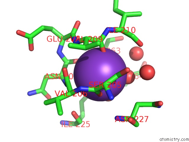
Mono view
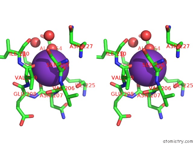
Stereo pair view
Mono view
Stereo pair view
A full contact list of Potassium with other atoms in the K binding
site number 1 of Human Type 1 Methionine Aminopeptidase in Complex with Ovalicin at 1.1 Ang within 5.0Å range:
|
Reference:
A.Addlagatta,
B.W.Matthews.
Structure of the Angiogenesis Inhibitor Ovalicin Bound to Its Noncognate Target, Human Type 1 Methionine Aminopeptidase. Protein Sci. V. 15 1842 2006.
ISSN: ISSN 0961-8368
PubMed: 16823043
DOI: 10.1110/PS.062278006
Page generated: Mon Aug 12 06:30:18 2024
ISSN: ISSN 0961-8368
PubMed: 16823043
DOI: 10.1110/PS.062278006
Last articles
Zn in 9J0NZn in 9J0O
Zn in 9J0P
Zn in 9FJX
Zn in 9EKB
Zn in 9C0F
Zn in 9CAH
Zn in 9CH0
Zn in 9CH3
Zn in 9CH1