Potassium »
PDB 2c44-2frz »
2feu »
Potassium in PDB 2feu: P450CAM From Pseudomonas Putida Reconstituted with Manganic Protoporphyrin IX
Enzymatic activity of P450CAM From Pseudomonas Putida Reconstituted with Manganic Protoporphyrin IX
All present enzymatic activity of P450CAM From Pseudomonas Putida Reconstituted with Manganic Protoporphyrin IX:
1.14.15.1;
1.14.15.1;
Protein crystallography data
The structure of P450CAM From Pseudomonas Putida Reconstituted with Manganic Protoporphyrin IX, PDB code: 2feu
was solved by
K.Von Koenig,
T.M.Makris,
S.G.Sligar,
I.Schlichting,
with X-Ray Crystallography technique. A brief refinement statistics is given in the table below:
| Resolution Low / High (Å) | 19.80 / 1.70 |
| Space group | P 1 21 1 |
| Cell size a, b, c (Å), α, β, γ (°) | 67.130, 62.230, 94.710, 90.00, 90.64, 90.00 |
| R / Rfree (%) | 21.4 / 23.8 |
Other elements in 2feu:
The structure of P450CAM From Pseudomonas Putida Reconstituted with Manganic Protoporphyrin IX also contains other interesting chemical elements:
| Manganese | (Mn) | 2 atoms |
Potassium Binding Sites:
The binding sites of Potassium atom in the P450CAM From Pseudomonas Putida Reconstituted with Manganic Protoporphyrin IX
(pdb code 2feu). This binding sites where shown within
5.0 Angstroms radius around Potassium atom.
In total 3 binding sites of Potassium where determined in the P450CAM From Pseudomonas Putida Reconstituted with Manganic Protoporphyrin IX, PDB code: 2feu:
Jump to Potassium binding site number: 1; 2; 3;
In total 3 binding sites of Potassium where determined in the P450CAM From Pseudomonas Putida Reconstituted with Manganic Protoporphyrin IX, PDB code: 2feu:
Jump to Potassium binding site number: 1; 2; 3;
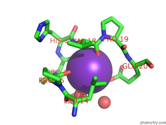
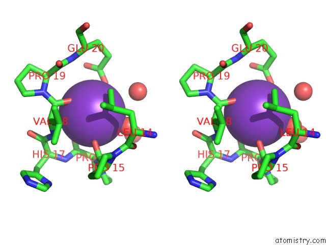
Potassium binding site 1 out of 3 in 2feu
Go back to
Potassium binding site 1 out
of 3 in the P450CAM From Pseudomonas Putida Reconstituted with Manganic Protoporphyrin IX
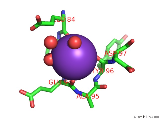
Mono view
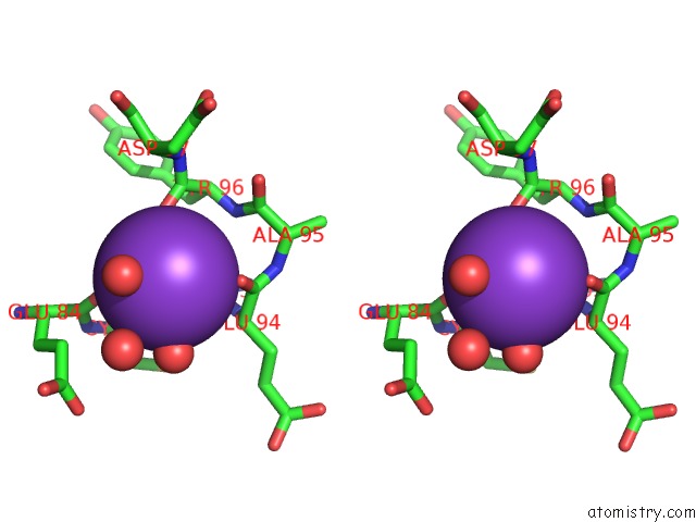
Stereo pair view
Mono view
Stereo pair view
A full contact list of Potassium with other atoms in the K binding
site number 1 of P450CAM From Pseudomonas Putida Reconstituted with Manganic Protoporphyrin IX within 5.0Å range:
|
Potassium binding site 2 out of 3 in 2feu
Go back to
Potassium binding site 2 out
of 3 in the P450CAM From Pseudomonas Putida Reconstituted with Manganic Protoporphyrin IX
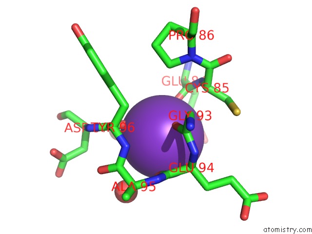
Mono view
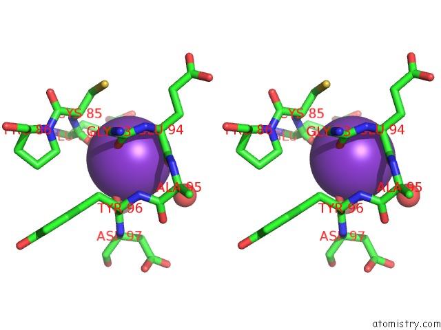
Stereo pair view
Mono view
Stereo pair view
A full contact list of Potassium with other atoms in the K binding
site number 2 of P450CAM From Pseudomonas Putida Reconstituted with Manganic Protoporphyrin IX within 5.0Å range:
|
Potassium binding site 3 out of 3 in 2feu
Go back to
Potassium binding site 3 out
of 3 in the P450CAM From Pseudomonas Putida Reconstituted with Manganic Protoporphyrin IX
Mono view
Stereo pair view
Mono view
Stereo pair view
A full contact list of Potassium with other atoms in the K binding
site number 3 of P450CAM From Pseudomonas Putida Reconstituted with Manganic Protoporphyrin IX within 5.0Å range:
|
Reference:
T.M.Makris,
K.Von Koenig,
I.Schlichting,
S.G.Sligar.
The Status of High-Valent Metal Oxo Complexes in the P450 Cytochromes. J.Inorg.Biochem. V. 100 507 2006.
ISSN: ISSN 0162-0134
PubMed: 16510191
DOI: 10.1016/J.JINORGBIO.2006.01.025
Page generated: Mon Aug 12 06:21:49 2024
ISSN: ISSN 0162-0134
PubMed: 16510191
DOI: 10.1016/J.JINORGBIO.2006.01.025
Last articles
Zn in 9J0NZn in 9J0O
Zn in 9J0P
Zn in 9FJX
Zn in 9EKB
Zn in 9C0F
Zn in 9CAH
Zn in 9CH0
Zn in 9CH3
Zn in 9CH1