Potassium »
PDB 2c44-2frz »
2fca »
Potassium in PDB 2fca: The Structure of Bstrmb
Enzymatic activity of The Structure of Bstrmb
All present enzymatic activity of The Structure of Bstrmb:
2.1.1.33;
2.1.1.33;
Protein crystallography data
The structure of The Structure of Bstrmb, PDB code: 2fca
was solved by
I.Zegers,
F.Van Vliet,
J.Bujnicki,
J.Kosinski,
D.Gigot,
L.Droogmans,
with X-Ray Crystallography technique. A brief refinement statistics is given in the table below:
| Resolution Low / High (Å) | 30.00 / 2.10 |
| Space group | H 3 |
| Cell size a, b, c (Å), α, β, γ (°) | 178.830, 178.830, 41.950, 90.00, 90.00, 120.00 |
| R / Rfree (%) | 20.2 / 23.3 |
Potassium Binding Sites:
The binding sites of Potassium atom in the The Structure of Bstrmb
(pdb code 2fca). This binding sites where shown within
5.0 Angstroms radius around Potassium atom.
In total 3 binding sites of Potassium where determined in the The Structure of Bstrmb, PDB code: 2fca:
Jump to Potassium binding site number: 1; 2; 3;
In total 3 binding sites of Potassium where determined in the The Structure of Bstrmb, PDB code: 2fca:
Jump to Potassium binding site number: 1; 2; 3;
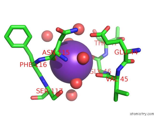
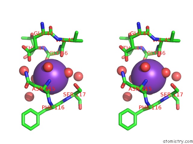
Potassium binding site 1 out of 3 in 2fca
Go back to
Potassium binding site 1 out
of 3 in the The Structure of Bstrmb
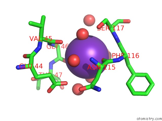
Mono view
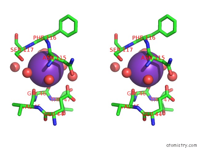
Stereo pair view
Mono view
Stereo pair view
A full contact list of Potassium with other atoms in the K binding
site number 1 of The Structure of Bstrmb within 5.0Å range:
|
Potassium binding site 2 out of 3 in 2fca
Go back to
Potassium binding site 2 out
of 3 in the The Structure of Bstrmb
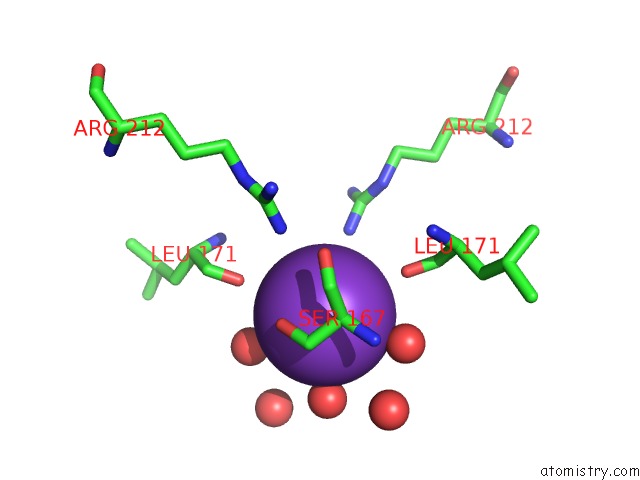
Mono view
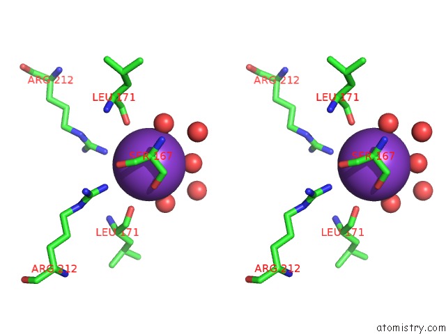
Stereo pair view
Mono view
Stereo pair view
A full contact list of Potassium with other atoms in the K binding
site number 2 of The Structure of Bstrmb within 5.0Å range:
|
Potassium binding site 3 out of 3 in 2fca
Go back to
Potassium binding site 3 out
of 3 in the The Structure of Bstrmb
Mono view
Stereo pair view
Mono view
Stereo pair view
A full contact list of Potassium with other atoms in the K binding
site number 3 of The Structure of Bstrmb within 5.0Å range:
|
Reference:
I.Zegers,
D.Gigot,
F.Van Vliet,
C.Tricot,
S.Aymerich,
J.M.Bujnicki,
J.Kosinski,
L.Droogmans.
Crystal Structure of Bacillus Subtilis Trmb, the Trna (M7G46) Methyltransferase. Nucleic Acids Res. V. 34 1925 2006.
ISSN: ISSN 0305-1048
PubMed: 16600901
DOI: 10.1093/NAR/GKL116
Page generated: Mon Aug 12 06:18:25 2024
ISSN: ISSN 0305-1048
PubMed: 16600901
DOI: 10.1093/NAR/GKL116
Last articles
Zn in 9JYWZn in 9IR4
Zn in 9IR3
Zn in 9GMX
Zn in 9GMW
Zn in 9JEJ
Zn in 9ERF
Zn in 9ERE
Zn in 9EGV
Zn in 9EGW