Potassium »
PDB 2c44-2frz »
2c44 »
Potassium in PDB 2c44: Crystal Structure of E. Coli Tryptophanase
Enzymatic activity of Crystal Structure of E. Coli Tryptophanase
All present enzymatic activity of Crystal Structure of E. Coli Tryptophanase:
4.1.99.1;
4.1.99.1;
Protein crystallography data
The structure of Crystal Structure of E. Coli Tryptophanase, PDB code: 2c44
was solved by
S.-Y.Ku,
P.Yip,
P.L.Howell,
with X-Ray Crystallography technique. A brief refinement statistics is given in the table below:
| Resolution Low / High (Å) | 500.00 / 2.81 |
| Space group | P 41 21 2 |
| Cell size a, b, c (Å), α, β, γ (°) | 215.510, 215.510, 107.560, 90.00, 90.00, 90.00 |
| R / Rfree (%) | 19.6 / 22 |
Potassium Binding Sites:
The binding sites of Potassium atom in the Crystal Structure of E. Coli Tryptophanase
(pdb code 2c44). This binding sites where shown within
5.0 Angstroms radius around Potassium atom.
In total 6 binding sites of Potassium where determined in the Crystal Structure of E. Coli Tryptophanase, PDB code: 2c44:
Jump to Potassium binding site number: 1; 2; 3; 4; 5; 6;
In total 6 binding sites of Potassium where determined in the Crystal Structure of E. Coli Tryptophanase, PDB code: 2c44:
Jump to Potassium binding site number: 1; 2; 3; 4; 5; 6;
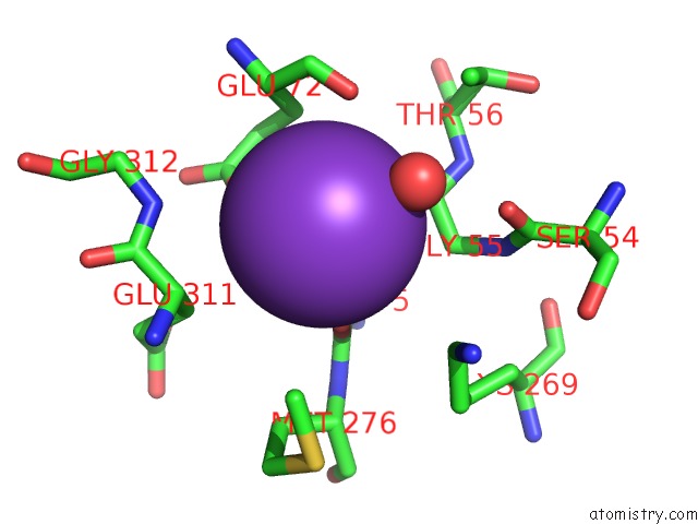
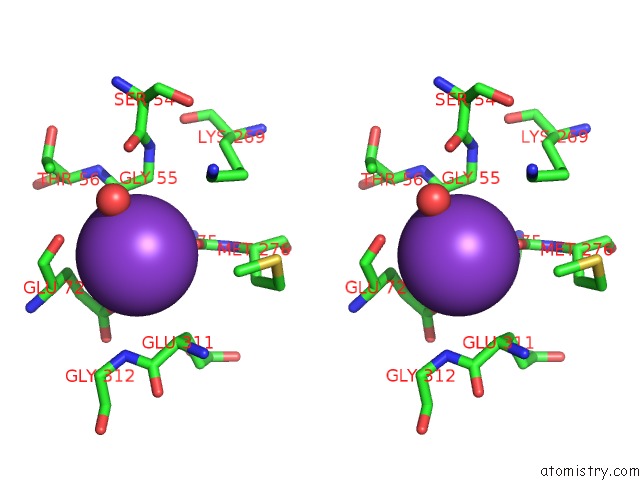
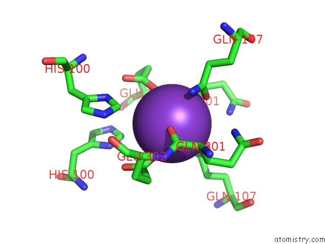
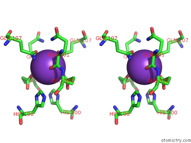
Potassium binding site 1 out of 6 in 2c44
Go back to
Potassium binding site 1 out
of 6 in the Crystal Structure of E. Coli Tryptophanase
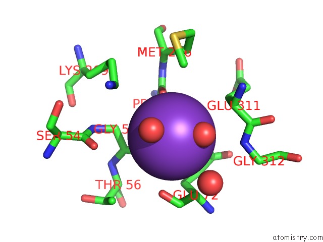
Mono view
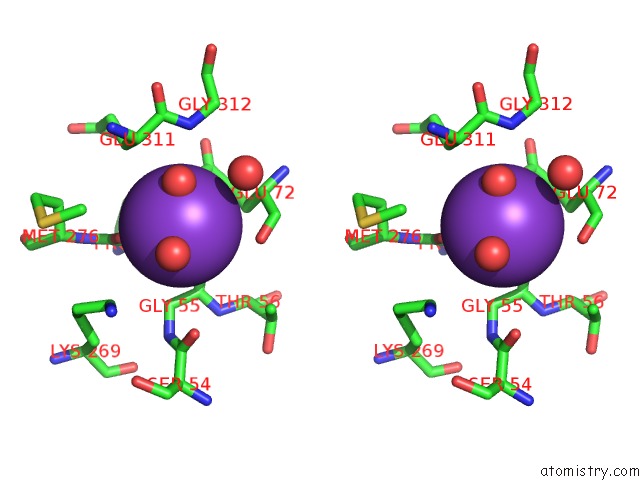
Stereo pair view
Mono view
Stereo pair view
A full contact list of Potassium with other atoms in the K binding
site number 1 of Crystal Structure of E. Coli Tryptophanase within 5.0Å range:
|
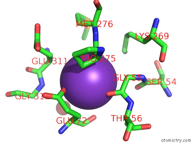
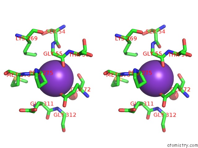
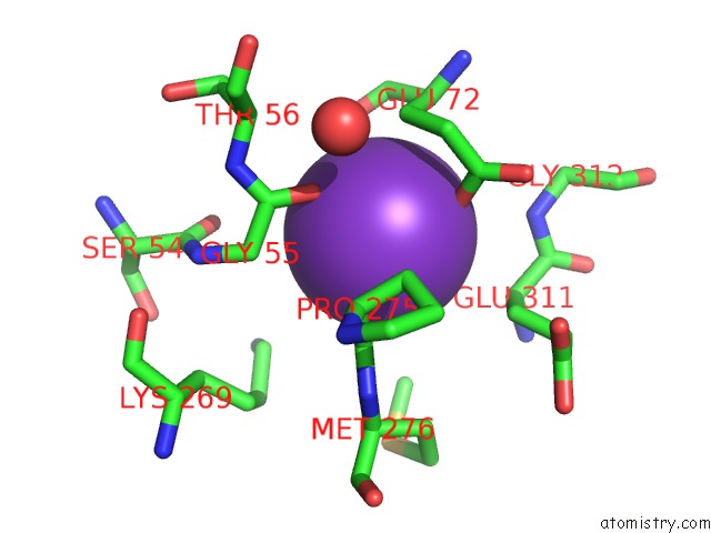
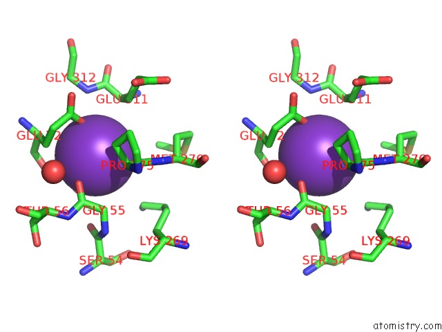
Potassium binding site 2 out of 6 in 2c44
Go back to
Potassium binding site 2 out
of 6 in the Crystal Structure of E. Coli Tryptophanase
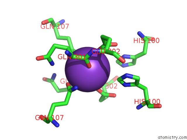
Mono view
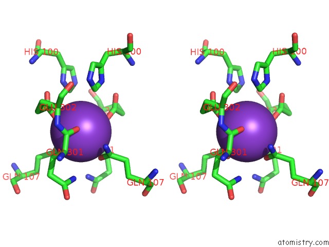
Stereo pair view
Mono view
Stereo pair view
A full contact list of Potassium with other atoms in the K binding
site number 2 of Crystal Structure of E. Coli Tryptophanase within 5.0Å range:
|
Potassium binding site 3 out of 6 in 2c44
Go back to
Potassium binding site 3 out
of 6 in the Crystal Structure of E. Coli Tryptophanase
Mono view
Stereo pair view
Mono view
Stereo pair view
A full contact list of Potassium with other atoms in the K binding
site number 3 of Crystal Structure of E. Coli Tryptophanase within 5.0Å range:
|
Potassium binding site 4 out of 6 in 2c44
Go back to
Potassium binding site 4 out
of 6 in the Crystal Structure of E. Coli Tryptophanase
Mono view
Stereo pair view
Mono view
Stereo pair view
A full contact list of Potassium with other atoms in the K binding
site number 4 of Crystal Structure of E. Coli Tryptophanase within 5.0Å range:
|
Potassium binding site 5 out of 6 in 2c44
Go back to
Potassium binding site 5 out
of 6 in the Crystal Structure of E. Coli Tryptophanase
Mono view
Stereo pair view
Mono view
Stereo pair view
A full contact list of Potassium with other atoms in the K binding
site number 5 of Crystal Structure of E. Coli Tryptophanase within 5.0Å range:
|
Potassium binding site 6 out of 6 in 2c44
Go back to
Potassium binding site 6 out
of 6 in the Crystal Structure of E. Coli Tryptophanase
Mono view
Stereo pair view
Mono view
Stereo pair view
A full contact list of Potassium with other atoms in the K binding
site number 6 of Crystal Structure of E. Coli Tryptophanase within 5.0Å range:
|
Reference:
S.-Y.Ku,
P.Yip,
P.L.Howell.
Structure of Escherichia Coli Tryptophanase Acta Crystallogr.,Sect.D V. 62 814 2006.
ISSN: ISSN 0907-4449
PubMed: 16790938
DOI: 10.1107/S0907444906019895
Page generated: Mon Aug 12 06:09:42 2024
ISSN: ISSN 0907-4449
PubMed: 16790938
DOI: 10.1107/S0907444906019895
Last articles
Zn in 9JYWZn in 9IR4
Zn in 9IR3
Zn in 9GMX
Zn in 9GMW
Zn in 9JEJ
Zn in 9ERF
Zn in 9ERE
Zn in 9EGV
Zn in 9EGW