Potassium »
PDB 2adp-2c13 »
2bup »
Potassium in PDB 2bup: T13G Mutant of the Atpase Fragment of Bovine HSC70
Protein crystallography data
The structure of T13G Mutant of the Atpase Fragment of Bovine HSC70, PDB code: 2bup
was solved by
M.C.Sousa,
D.B.Mckay,
with X-Ray Crystallography technique. A brief refinement statistics is given in the table below:
| Resolution Low / High (Å) | 30.00 / 1.70 |
| Space group | P 21 21 21 |
| Cell size a, b, c (Å), α, β, γ (°) | 143.350, 64.300, 46.230, 90.00, 90.00, 90.00 |
| R / Rfree (%) | 19.5 / 22.4 |
Other elements in 2bup:
The structure of T13G Mutant of the Atpase Fragment of Bovine HSC70 also contains other interesting chemical elements:
| Magnesium | (Mg) | 1 atom |
| Chlorine | (Cl) | 2 atoms |
Potassium Binding Sites:
The binding sites of Potassium atom in the T13G Mutant of the Atpase Fragment of Bovine HSC70
(pdb code 2bup). This binding sites where shown within
5.0 Angstroms radius around Potassium atom.
In total 2 binding sites of Potassium where determined in the T13G Mutant of the Atpase Fragment of Bovine HSC70, PDB code: 2bup:
Jump to Potassium binding site number: 1; 2;
In total 2 binding sites of Potassium where determined in the T13G Mutant of the Atpase Fragment of Bovine HSC70, PDB code: 2bup:
Jump to Potassium binding site number: 1; 2;
Potassium binding site 1 out of 2 in 2bup
Go back to
Potassium binding site 1 out
of 2 in the T13G Mutant of the Atpase Fragment of Bovine HSC70
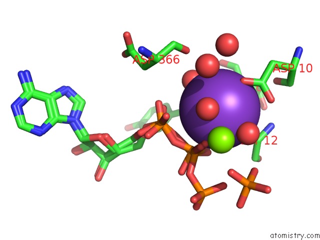
Mono view
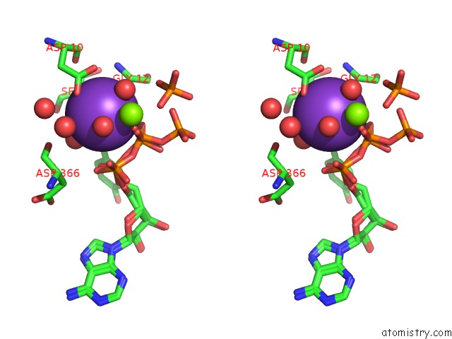
Stereo pair view
Mono view
Stereo pair view
A full contact list of Potassium with other atoms in the K binding
site number 1 of T13G Mutant of the Atpase Fragment of Bovine HSC70 within 5.0Å range:
|
Potassium binding site 2 out of 2 in 2bup
Go back to
Potassium binding site 2 out
of 2 in the T13G Mutant of the Atpase Fragment of Bovine HSC70
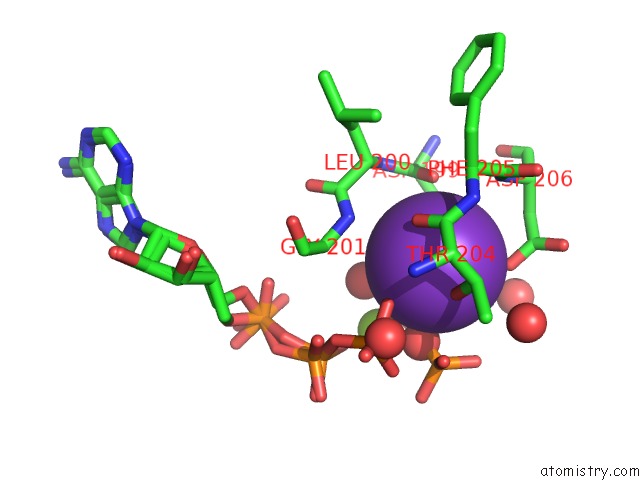
Mono view
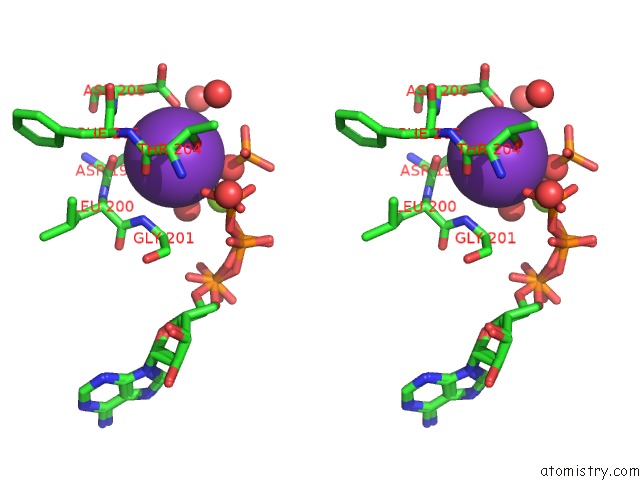
Stereo pair view
Mono view
Stereo pair view
A full contact list of Potassium with other atoms in the K binding
site number 2 of T13G Mutant of the Atpase Fragment of Bovine HSC70 within 5.0Å range:
|
Reference:
M.C.Sousa,
D.B.Mckay.
The Hydroxyl of Threonine 13 of the Bovine 70-kDa Heat Shock Cognate Protein Is Essential For Transducing the Atp-Induced Conformational Change. Biochemistry V. 37 15392 1998.
ISSN: ISSN 0006-2960
PubMed: 9799500
DOI: 10.1021/BI981510X
Page generated: Mon Aug 12 06:08:39 2024
ISSN: ISSN 0006-2960
PubMed: 9799500
DOI: 10.1021/BI981510X
Last articles
Zn in 9J0NZn in 9J0O
Zn in 9J0P
Zn in 9FJX
Zn in 9EKB
Zn in 9C0F
Zn in 9CAH
Zn in 9CH0
Zn in 9CH3
Zn in 9CH1