Potassium »
PDB 2adp-2c13 »
2b3l »
Potassium in PDB 2b3l: Crystal Structure of Type I Human Methionine Aminopeptidase in the Apo Form
Enzymatic activity of Crystal Structure of Type I Human Methionine Aminopeptidase in the Apo Form
All present enzymatic activity of Crystal Structure of Type I Human Methionine Aminopeptidase in the Apo Form:
3.4.11.18;
3.4.11.18;
Protein crystallography data
The structure of Crystal Structure of Type I Human Methionine Aminopeptidase in the Apo Form, PDB code: 2b3l
was solved by
A.Addlagatta,
X.Hu,
J.O.Liu,
B.W.Matthews,
with X-Ray Crystallography technique. A brief refinement statistics is given in the table below:
| Resolution Low / High (Å) | 50.00 / 1.50 |
| Space group | P 1 21 1 |
| Cell size a, b, c (Å), α, β, γ (°) | 47.217, 77.276, 47.850, 90.00, 91.59, 90.00 |
| R / Rfree (%) | 11.1 / 18 |
Potassium Binding Sites:
The binding sites of Potassium atom in the Crystal Structure of Type I Human Methionine Aminopeptidase in the Apo Form
(pdb code 2b3l). This binding sites where shown within
5.0 Angstroms radius around Potassium atom.
In total only one binding site of Potassium was determined in the Crystal Structure of Type I Human Methionine Aminopeptidase in the Apo Form, PDB code: 2b3l:
In total only one binding site of Potassium was determined in the Crystal Structure of Type I Human Methionine Aminopeptidase in the Apo Form, PDB code: 2b3l:
Potassium binding site 1 out of 1 in 2b3l
Go back to
Potassium binding site 1 out
of 1 in the Crystal Structure of Type I Human Methionine Aminopeptidase in the Apo Form
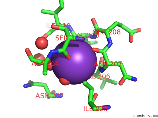
Mono view
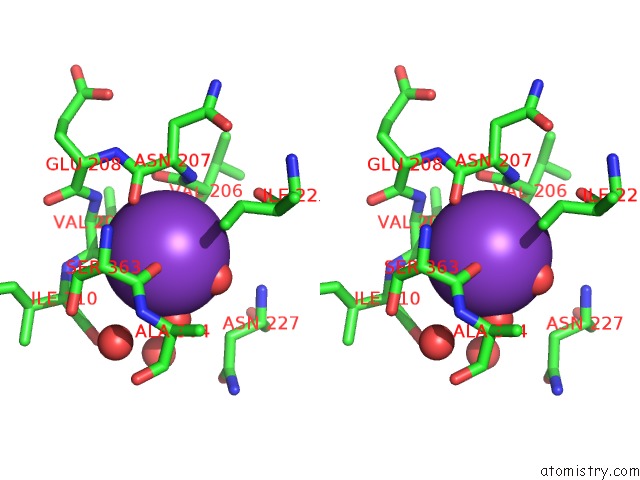
Stereo pair view
Mono view
Stereo pair view
A full contact list of Potassium with other atoms in the K binding
site number 1 of Crystal Structure of Type I Human Methionine Aminopeptidase in the Apo Form within 5.0Å range:
|
Reference:
A.Addlagatta,
X.Hu,
J.O.Liu,
B.W.Matthews.
Structural Basis For the Functional Differences Between Type I and Type II Human Methionine Aminopeptidases(,). Biochemistry V. 44 14741 2005.
ISSN: ISSN 0006-2960
PubMed: 16274222
DOI: 10.1021/BI051691K
Page generated: Mon Aug 12 06:05:12 2024
ISSN: ISSN 0006-2960
PubMed: 16274222
DOI: 10.1021/BI051691K
Last articles
Zn in 9JYWZn in 9IR4
Zn in 9IR3
Zn in 9GMX
Zn in 9GMW
Zn in 9JEJ
Zn in 9ERF
Zn in 9ERE
Zn in 9EGV
Zn in 9EGW