Potassium »
PDB 1yjn-2aaq »
2a6n »
Potassium in PDB 2a6n: Dihydrodipicolinate Synthase (E. Coli)- Mutant R138A
Enzymatic activity of Dihydrodipicolinate Synthase (E. Coli)- Mutant R138A
All present enzymatic activity of Dihydrodipicolinate Synthase (E. Coli)- Mutant R138A:
4.2.1.52;
4.2.1.52;
Protein crystallography data
The structure of Dihydrodipicolinate Synthase (E. Coli)- Mutant R138A, PDB code: 2a6n
was solved by
R.C.Dobson,
S.R.Devenish,
L.A.Turner,
V.R.Clifford,
F.G.Pearce,
G.B.Jameson,
J.A.Gerrard,
with X-Ray Crystallography technique. A brief refinement statistics is given in the table below:
| Resolution Low / High (Å) | 33.33 / 1.94 |
| Space group | P 31 2 1 |
| Cell size a, b, c (Å), α, β, γ (°) | 120.989, 120.989, 111.302, 90.00, 90.00, 120.00 |
| R / Rfree (%) | 16.3 / 19.1 |
Potassium Binding Sites:
The binding sites of Potassium atom in the Dihydrodipicolinate Synthase (E. Coli)- Mutant R138A
(pdb code 2a6n). This binding sites where shown within
5.0 Angstroms radius around Potassium atom.
In total 2 binding sites of Potassium where determined in the Dihydrodipicolinate Synthase (E. Coli)- Mutant R138A, PDB code: 2a6n:
Jump to Potassium binding site number: 1; 2;
In total 2 binding sites of Potassium where determined in the Dihydrodipicolinate Synthase (E. Coli)- Mutant R138A, PDB code: 2a6n:
Jump to Potassium binding site number: 1; 2;
Potassium binding site 1 out of 2 in 2a6n
Go back to
Potassium binding site 1 out
of 2 in the Dihydrodipicolinate Synthase (E. Coli)- Mutant R138A
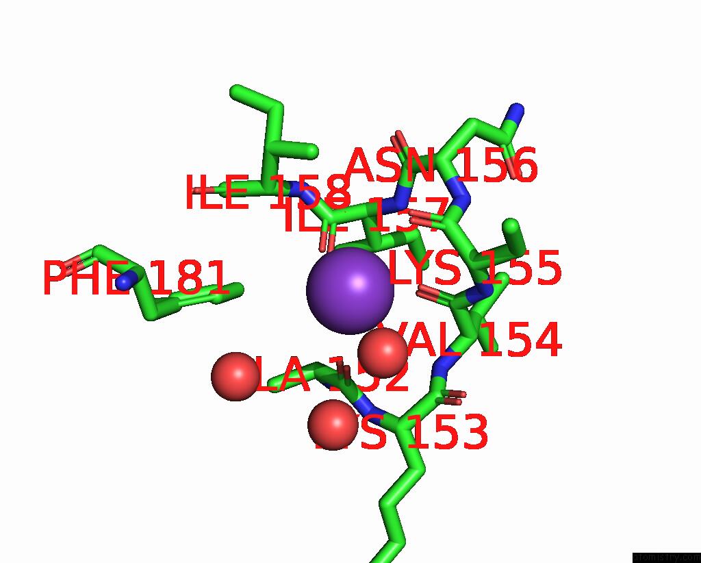
Mono view
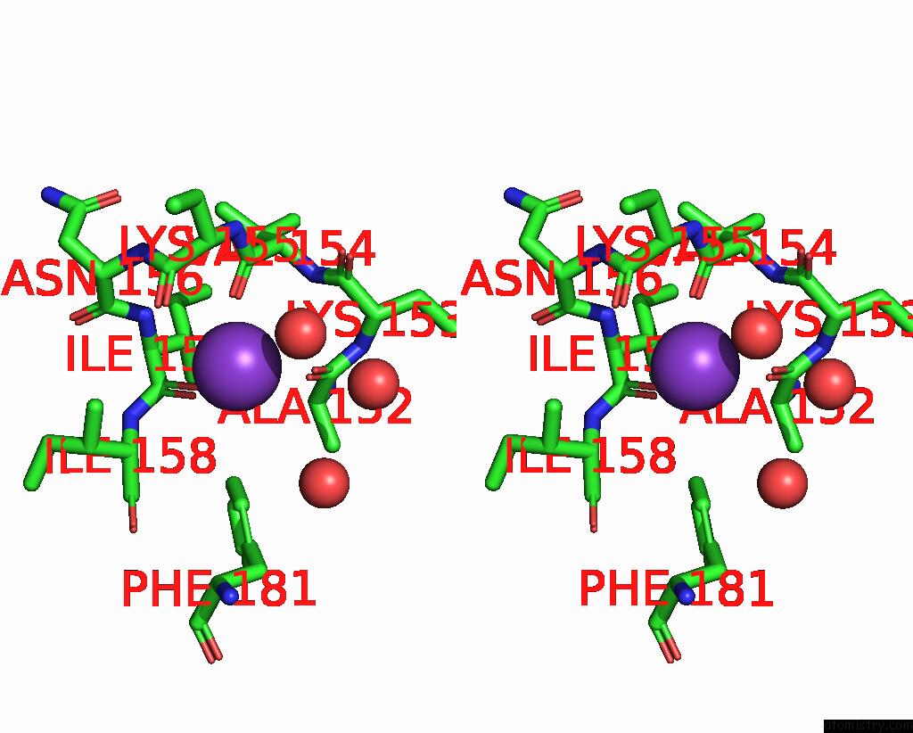
Stereo pair view
Mono view
Stereo pair view
A full contact list of Potassium with other atoms in the K binding
site number 1 of Dihydrodipicolinate Synthase (E. Coli)- Mutant R138A within 5.0Å range:
|
Potassium binding site 2 out of 2 in 2a6n
Go back to
Potassium binding site 2 out
of 2 in the Dihydrodipicolinate Synthase (E. Coli)- Mutant R138A

Mono view
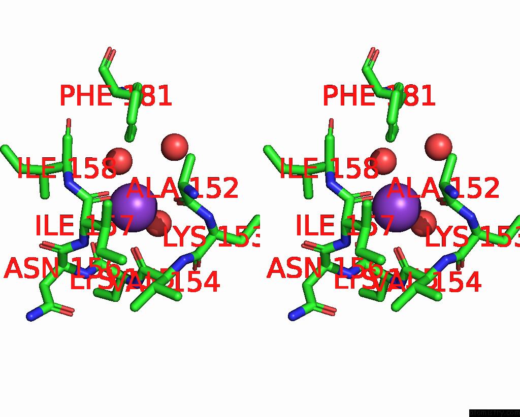
Stereo pair view
Mono view
Stereo pair view
A full contact list of Potassium with other atoms in the K binding
site number 2 of Dihydrodipicolinate Synthase (E. Coli)- Mutant R138A within 5.0Å range:
|
Reference:
R.C.Dobson,
S.R.Devenish,
L.A.Turner,
V.R.Clifford,
F.G.Pearce,
G.B.Jameson,
J.A.Gerrard.
Role of Arginine 138 in the Catalysis and Regulation of Escherichia Coli Dihydrodipicolinate Synthase. Biochemistry V. 44 13007 2005.
ISSN: ISSN 0006-2960
PubMed: 16185069
DOI: 10.1021/BI051281W
Page generated: Mon Aug 12 05:59:26 2024
ISSN: ISSN 0006-2960
PubMed: 16185069
DOI: 10.1021/BI051281W
Last articles
Zn in 9J0NZn in 9J0O
Zn in 9J0P
Zn in 9FJX
Zn in 9EKB
Zn in 9C0F
Zn in 9CAH
Zn in 9CH0
Zn in 9CH3
Zn in 9CH1