Potassium in PDB 1zho: The Structure of A Ribosomal Protein L1 in Complex with Mrna
Protein crystallography data
The structure of The Structure of A Ribosomal Protein L1 in Complex with Mrna, PDB code: 1zho
was solved by
N.Nevskaya,
S.Tishchenko,
S.Volchkov,
V.Kljashtorny,
E.Nikonova,
O.Nikonov,
A.Nikulin,
C.Kohrer,
W.Piendl,
R.Zimmermann,
P.Stockley,
M.Garber,
S.Nikonov,
with X-Ray Crystallography technique. A brief refinement statistics is given in the table below:
Resolution Low / High (Å)
|
37.06 /
2.60
|
Space group
|
P 1 21 1
|
Cell size a, b, c (Å), α, β, γ (°)
|
71.600,
101.900,
119.600,
90.00,
93.66,
90.00
|
R / Rfree (%)
|
21.8 /
28.1
|
Potassium Binding Sites:
The binding sites of Potassium atom in the The Structure of A Ribosomal Protein L1 in Complex with Mrna
(pdb code 1zho). This binding sites where shown within
5.0 Angstroms radius around Potassium atom.
In total 4 binding sites of Potassium where determined in the
The Structure of A Ribosomal Protein L1 in Complex with Mrna, PDB code: 1zho:
Jump to Potassium binding site number:
1;
2;
3;
4;
Potassium binding site 1 out
of 4 in 1zho
Go back to
Potassium Binding Sites List in 1zho
Potassium binding site 1 out
of 4 in the The Structure of A Ribosomal Protein L1 in Complex with Mrna
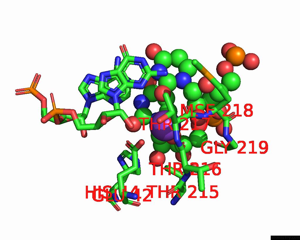
Mono view
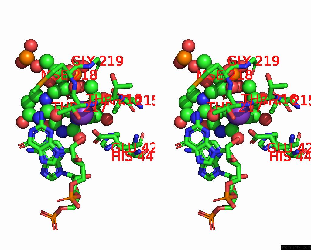
Stereo pair view
|
A full contact list of Potassium with other atoms in the K binding
site number 1 of The Structure of A Ribosomal Protein L1 in Complex with Mrna within 5.0Å range:
probe
|
atom
|
residue
|
distance (Å)
|
B
|
Occ
|
D:K39
b:42.0
occ:1.00
|
O
|
C:THR216
|
2.9
|
45.3
|
1.0
|
O2
|
D:U30
|
3.0
|
45.7
|
1.0
|
O
|
C:THR217
|
3.1
|
43.3
|
1.0
|
O2'
|
D:U30
|
3.3
|
38.8
|
1.0
|
OE2
|
C:GLU42
|
3.3
|
42.6
|
1.0
|
O4'
|
D:U31
|
3.4
|
40.9
|
1.0
|
CD
|
C:GLU42
|
3.6
|
44.9
|
1.0
|
C
|
C:THR217
|
3.6
|
43.5
|
1.0
|
OE1
|
C:GLU42
|
3.7
|
48.7
|
1.0
|
CA
|
C:THR217
|
3.8
|
42.9
|
1.0
|
C
|
C:THR216
|
3.8
|
44.4
|
1.0
|
CG2
|
C:THR215
|
3.8
|
39.3
|
1.0
|
C2'
|
D:U30
|
3.9
|
37.9
|
1.0
|
C1'
|
D:U30
|
4.0
|
38.7
|
1.0
|
C1'
|
D:U31
|
4.0
|
40.0
|
1.0
|
C2
|
D:U30
|
4.1
|
43.9
|
1.0
|
N
|
C:THR217
|
4.2
|
44.1
|
1.0
|
N1
|
D:U31
|
4.4
|
39.6
|
1.0
|
N3
|
D:G10
|
4.4
|
39.9
|
1.0
|
OG1
|
C:THR215
|
4.5
|
40.8
|
1.0
|
C4'
|
D:U31
|
4.6
|
40.6
|
1.0
|
N1
|
D:U30
|
4.6
|
41.4
|
1.0
|
CG
|
C:GLU42
|
4.6
|
43.8
|
1.0
|
C2
|
D:A9
|
4.7
|
37.9
|
1.0
|
NE2
|
C:HIS44
|
4.7
|
45.7
|
1.0
|
O
|
C:GLY219
|
4.7
|
46.3
|
1.0
|
N
|
C:MSE218
|
4.7
|
44.9
|
1.0
|
C6
|
D:U31
|
4.7
|
37.9
|
1.0
|
CB
|
C:THR215
|
4.8
|
41.0
|
1.0
|
C1'
|
D:G10
|
4.9
|
37.9
|
1.0
|
N2
|
D:G10
|
4.9
|
42.2
|
1.0
|
C2
|
D:U31
|
4.9
|
39.7
|
1.0
|
C5'
|
D:U31
|
4.9
|
38.3
|
1.0
|
O2'
|
D:G10
|
4.9
|
36.9
|
1.0
|
N
|
C:THR216
|
5.0
|
43.5
|
1.0
|
|
Potassium binding site 2 out
of 4 in 1zho
Go back to
Potassium Binding Sites List in 1zho
Potassium binding site 2 out
of 4 in the The Structure of A Ribosomal Protein L1 in Complex with Mrna
Mono view
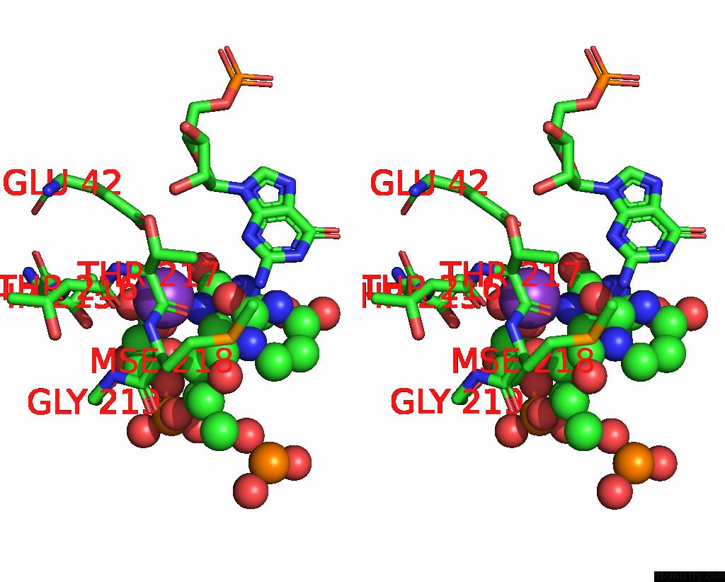
Stereo pair view
|
A full contact list of Potassium with other atoms in the K binding
site number 2 of The Structure of A Ribosomal Protein L1 in Complex with Mrna within 5.0Å range:
probe
|
atom
|
residue
|
distance (Å)
|
B
|
Occ
|
H:K39
b:61.1
occ:1.00
|
O2'
|
H:U30
|
2.6
|
42.2
|
1.0
|
O
|
G:THR216
|
2.6
|
51.4
|
1.0
|
O2
|
H:U30
|
3.0
|
45.6
|
1.0
|
O
|
G:THR217
|
3.0
|
50.6
|
1.0
|
O4'
|
H:U31
|
3.2
|
45.0
|
1.0
|
C2'
|
H:U30
|
3.4
|
41.8
|
1.0
|
OE1
|
G:GLU42
|
3.5
|
32.5
|
1.0
|
C
|
G:THR217
|
3.5
|
51.0
|
1.0
|
C
|
G:THR216
|
3.5
|
48.9
|
1.0
|
C1'
|
H:U30
|
3.6
|
43.2
|
1.0
|
CA
|
G:THR217
|
3.7
|
50.6
|
1.0
|
OE2
|
G:GLU42
|
3.8
|
32.1
|
1.0
|
OG1
|
G:THR215
|
3.8
|
46.4
|
1.0
|
CD
|
G:GLU42
|
3.9
|
32.4
|
1.0
|
N
|
G:THR217
|
4.0
|
49.7
|
1.0
|
C2
|
H:U30
|
4.0
|
45.7
|
1.0
|
C4'
|
H:U31
|
4.2
|
44.2
|
1.0
|
CG2
|
G:THR215
|
4.2
|
45.4
|
1.0
|
C1'
|
H:U31
|
4.2
|
45.6
|
1.0
|
C5'
|
H:U31
|
4.2
|
44.3
|
1.0
|
N1
|
H:U30
|
4.3
|
45.2
|
1.0
|
O
|
G:GLY219
|
4.4
|
51.9
|
1.0
|
N
|
G:MSE218
|
4.5
|
53.0
|
1.0
|
N
|
G:THR216
|
4.6
|
45.4
|
1.0
|
CB
|
G:THR215
|
4.6
|
46.0
|
1.0
|
N1
|
H:U31
|
4.6
|
47.8
|
1.0
|
C6
|
H:U31
|
4.7
|
48.3
|
1.0
|
O4'
|
H:U30
|
4.7
|
41.0
|
1.0
|
O5'
|
H:U31
|
4.7
|
45.2
|
1.0
|
CA
|
G:THR216
|
4.8
|
46.7
|
1.0
|
C3'
|
H:U30
|
4.8
|
42.0
|
1.0
|
N3
|
H:G10
|
4.9
|
47.2
|
1.0
|
N2
|
H:G10
|
5.0
|
46.9
|
1.0
|
|
Potassium binding site 3 out
of 4 in 1zho
Go back to
Potassium Binding Sites List in 1zho
Potassium binding site 3 out
of 4 in the The Structure of A Ribosomal Protein L1 in Complex with Mrna
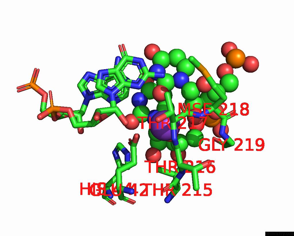
Mono view
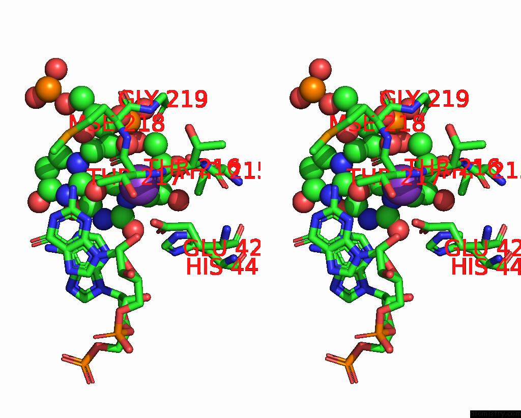
Stereo pair view
|
A full contact list of Potassium with other atoms in the K binding
site number 3 of The Structure of A Ribosomal Protein L1 in Complex with Mrna within 5.0Å range:
probe
|
atom
|
residue
|
distance (Å)
|
B
|
Occ
|
A:K229
b:33.2
occ:1.00
|
O
|
A:THR216
|
2.7
|
36.1
|
1.0
|
O2
|
B:U30
|
3.1
|
37.6
|
1.0
|
O2'
|
B:U30
|
3.1
|
33.3
|
1.0
|
O
|
A:THR217
|
3.2
|
38.1
|
1.0
|
O4'
|
B:U31
|
3.3
|
30.0
|
1.0
|
OE2
|
A:GLU42
|
3.4
|
28.7
|
1.0
|
OE1
|
A:GLU42
|
3.5
|
34.1
|
1.0
|
CD
|
A:GLU42
|
3.5
|
31.5
|
1.0
|
C
|
A:THR216
|
3.7
|
34.1
|
1.0
|
C
|
A:THR217
|
3.7
|
37.8
|
1.0
|
C2'
|
B:U30
|
3.8
|
34.3
|
1.0
|
OG1
|
A:THR215
|
3.9
|
34.8
|
1.0
|
CA
|
A:THR217
|
3.9
|
35.6
|
1.0
|
C1'
|
B:U31
|
4.0
|
30.9
|
1.0
|
C1'
|
B:U30
|
4.0
|
35.5
|
1.0
|
C2
|
B:U30
|
4.2
|
38.0
|
1.0
|
CG2
|
A:THR215
|
4.2
|
32.6
|
1.0
|
N
|
A:THR217
|
4.3
|
34.9
|
1.0
|
N1
|
B:U31
|
4.4
|
32.8
|
1.0
|
C4'
|
B:U31
|
4.4
|
30.8
|
1.0
|
CG
|
A:GLU42
|
4.5
|
30.0
|
1.0
|
N3
|
B:G10
|
4.6
|
36.6
|
1.0
|
O
|
A:GLY219
|
4.6
|
35.0
|
1.0
|
N1
|
B:U30
|
4.6
|
37.4
|
1.0
|
CB
|
A:THR215
|
4.7
|
33.3
|
1.0
|
N
|
A:MSE218
|
4.7
|
41.0
|
1.0
|
C6
|
B:U31
|
4.7
|
32.9
|
1.0
|
N2
|
B:G10
|
4.7
|
37.3
|
1.0
|
N
|
A:THR216
|
4.8
|
32.2
|
1.0
|
C5'
|
B:U31
|
4.8
|
31.8
|
1.0
|
C2
|
B:A9
|
4.9
|
29.6
|
1.0
|
NE2
|
A:HIS44
|
4.9
|
31.9
|
1.0
|
CA
|
A:THR216
|
4.9
|
33.4
|
1.0
|
|
Potassium binding site 4 out
of 4 in 1zho
Go back to
Potassium Binding Sites List in 1zho
Potassium binding site 4 out
of 4 in the The Structure of A Ribosomal Protein L1 in Complex with Mrna
Mono view
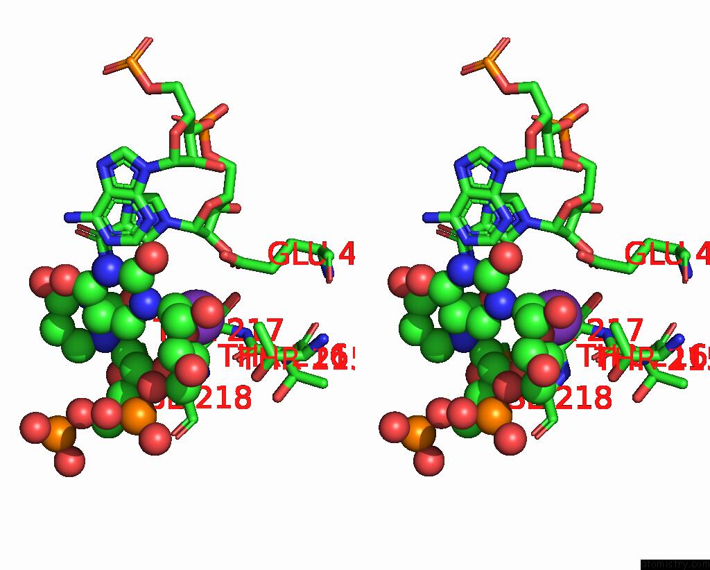
Stereo pair view
|
A full contact list of Potassium with other atoms in the K binding
site number 4 of The Structure of A Ribosomal Protein L1 in Complex with Mrna within 5.0Å range:
probe
|
atom
|
residue
|
distance (Å)
|
B
|
Occ
|
E:K229
b:66.4
occ:1.00
|
O2'
|
F:U30
|
3.0
|
55.3
|
1.0
|
O
|
E:THR216
|
3.1
|
58.3
|
1.0
|
O
|
E:THR217
|
3.2
|
56.5
|
1.0
|
O4'
|
F:U31
|
3.2
|
49.1
|
1.0
|
O2
|
F:U30
|
3.2
|
60.4
|
1.0
|
OE1
|
E:GLU42
|
3.4
|
42.9
|
1.0
|
OE2
|
E:GLU42
|
3.5
|
45.4
|
1.0
|
CD
|
E:GLU42
|
3.5
|
44.4
|
1.0
|
C
|
E:THR217
|
3.6
|
57.9
|
1.0
|
CA
|
E:THR217
|
3.7
|
57.4
|
1.0
|
CG2
|
E:THR215
|
3.7
|
57.4
|
1.0
|
C
|
E:THR216
|
3.8
|
57.5
|
1.0
|
C2'
|
F:U30
|
3.8
|
52.6
|
1.0
|
C1'
|
F:U30
|
3.9
|
54.0
|
1.0
|
C1'
|
F:U31
|
4.0
|
50.2
|
1.0
|
N
|
E:THR217
|
4.1
|
57.6
|
1.0
|
C2
|
F:U30
|
4.3
|
58.1
|
1.0
|
C4'
|
F:U31
|
4.3
|
49.3
|
1.0
|
N1
|
F:U31
|
4.3
|
52.1
|
1.0
|
OG1
|
E:THR215
|
4.3
|
57.9
|
1.0
|
CG
|
E:GLU42
|
4.4
|
43.9
|
1.0
|
C6
|
F:U31
|
4.4
|
51.9
|
1.0
|
C5'
|
F:U31
|
4.5
|
50.0
|
1.0
|
N
|
E:MSE218
|
4.6
|
58.9
|
1.0
|
N1
|
F:U30
|
4.7
|
56.7
|
1.0
|
CB
|
E:THR215
|
4.7
|
57.0
|
1.0
|
N3
|
F:G10
|
4.8
|
59.5
|
1.0
|
O5'
|
F:U31
|
4.8
|
51.8
|
1.0
|
C2
|
F:A9
|
4.9
|
51.4
|
1.0
|
N
|
E:THR216
|
4.9
|
55.0
|
1.0
|
CA
|
E:THR216
|
5.0
|
57.1
|
1.0
|
|
Reference:
N.Nevskaya,
S.Tishchenko,
S.Volchkov,
V.Kljashtorny,
E.Nikonova,
O.Nikonov,
A.Nikulin,
C.Kohrer,
W.Piendl,
R.Zimmermann,
P.Stockley,
M.Garber,
S.Nikonov.
New Insights Into the Interaction of Ribosomal Protein L1 with Rna. J.Mol.Biol. V. 355 747 2006.
ISSN: ISSN 0022-2836
PubMed: 16330048
DOI: 10.1016/J.JMB.2005.10.084
Page generated: Mon Aug 12 05:53:51 2024
|