Potassium »
PDB 1yjn-2aaq »
1z3z »
Potassium in PDB 1z3z: The Crystal Structure of A Dgd Mutant: Q52A
Enzymatic activity of The Crystal Structure of A Dgd Mutant: Q52A
All present enzymatic activity of The Crystal Structure of A Dgd Mutant: Q52A:
4.1.1.64;
4.1.1.64;
Protein crystallography data
The structure of The Crystal Structure of A Dgd Mutant: Q52A, PDB code: 1z3z
was solved by
E.J.Fogle,
W.Liu,
M.D.Toney,
with X-Ray Crystallography technique. A brief refinement statistics is given in the table below:
| Resolution Low / High (Å) | 50.00 / 2.90 |
| Space group | P 64 2 2 |
| Cell size a, b, c (Å), α, β, γ (°) | 150.040, 150.040, 84.420, 90.00, 90.00, 120.00 |
| R / Rfree (%) | 20.9 / 26.6 |
Other elements in 1z3z:
The structure of The Crystal Structure of A Dgd Mutant: Q52A also contains other interesting chemical elements:
| Sodium | (Na) | 1 atom |
Potassium Binding Sites:
The binding sites of Potassium atom in the The Crystal Structure of A Dgd Mutant: Q52A
(pdb code 1z3z). This binding sites where shown within
5.0 Angstroms radius around Potassium atom.
In total only one binding site of Potassium was determined in the The Crystal Structure of A Dgd Mutant: Q52A, PDB code: 1z3z:
In total only one binding site of Potassium was determined in the The Crystal Structure of A Dgd Mutant: Q52A, PDB code: 1z3z:
Potassium binding site 1 out of 1 in 1z3z
Go back to
Potassium binding site 1 out
of 1 in the The Crystal Structure of A Dgd Mutant: Q52A

Mono view
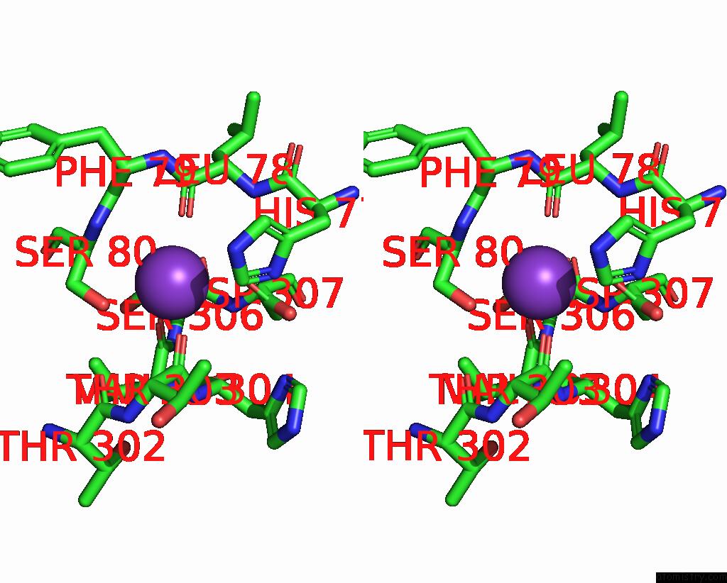
Stereo pair view
Mono view
Stereo pair view
A full contact list of Potassium with other atoms in the K binding
site number 1 of The Crystal Structure of A Dgd Mutant: Q52A within 5.0Å range:
|
Reference:
E.J.Fogle,
W.Liu,
S.T.Woon,
J.W.Keller,
M.D.Toney.
Role of Q52 in Catalysis of Decarboxylation and Transamination in Dialkylglycine Decarboxylase. Biochemistry V. 44 16392 2005.
ISSN: ISSN 0006-2960
PubMed: 16342932
DOI: 10.1021/BI051475B
Page generated: Mon Aug 12 05:53:17 2024
ISSN: ISSN 0006-2960
PubMed: 16342932
DOI: 10.1021/BI051475B
Last articles
Zn in 9J0NZn in 9J0O
Zn in 9J0P
Zn in 9FJX
Zn in 9EKB
Zn in 9C0F
Zn in 9CAH
Zn in 9CH0
Zn in 9CH3
Zn in 9CH1