Potassium »
PDB 1w2b-1yj9 »
1w85 »
Potassium in PDB 1w85: The Crystal Structure of Pyruvate Dehydrogenase E1 Bound to the Peripheral Subunit Binding Domain of E2
Enzymatic activity of The Crystal Structure of Pyruvate Dehydrogenase E1 Bound to the Peripheral Subunit Binding Domain of E2
All present enzymatic activity of The Crystal Structure of Pyruvate Dehydrogenase E1 Bound to the Peripheral Subunit Binding Domain of E2:
1.2.4.1; 2.3.1.12;
1.2.4.1; 2.3.1.12;
Protein crystallography data
The structure of The Crystal Structure of Pyruvate Dehydrogenase E1 Bound to the Peripheral Subunit Binding Domain of E2, PDB code: 1w85
was solved by
R.A.W.Frank,
J.V.Pratap,
X.Y.Pei,
R.N.Perham,
B.F.Luisi,
with X-Ray Crystallography technique. A brief refinement statistics is given in the table below:
| Resolution Low / High (Å) | 20.00 / 2.00 |
| Space group | P 1 21 1 |
| Cell size a, b, c (Å), α, β, γ (°) | 68.270, 232.330, 91.924, 90.00, 90.81, 90.00 |
| R / Rfree (%) | 17.6 / 21.5 |
Other elements in 1w85:
The structure of The Crystal Structure of Pyruvate Dehydrogenase E1 Bound to the Peripheral Subunit Binding Domain of E2 also contains other interesting chemical elements:
| Magnesium | (Mg) | 6 atoms |
Potassium Binding Sites:
The binding sites of Potassium atom in the The Crystal Structure of Pyruvate Dehydrogenase E1 Bound to the Peripheral Subunit Binding Domain of E2
(pdb code 1w85). This binding sites where shown within
5.0 Angstroms radius around Potassium atom.
In total 4 binding sites of Potassium where determined in the The Crystal Structure of Pyruvate Dehydrogenase E1 Bound to the Peripheral Subunit Binding Domain of E2, PDB code: 1w85:
Jump to Potassium binding site number: 1; 2; 3; 4;
In total 4 binding sites of Potassium where determined in the The Crystal Structure of Pyruvate Dehydrogenase E1 Bound to the Peripheral Subunit Binding Domain of E2, PDB code: 1w85:
Jump to Potassium binding site number: 1; 2; 3; 4;
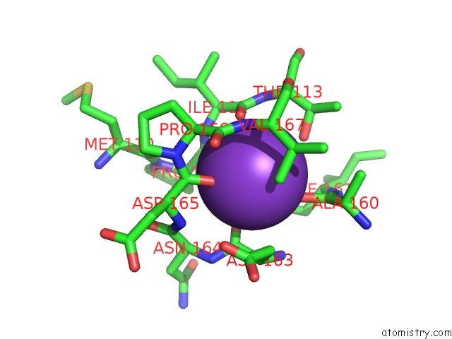
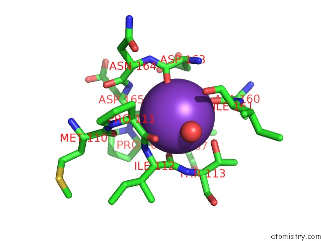
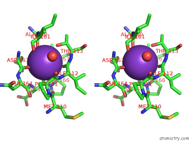
Potassium binding site 1 out of 4 in 1w85
Go back to
Potassium binding site 1 out
of 4 in the The Crystal Structure of Pyruvate Dehydrogenase E1 Bound to the Peripheral Subunit Binding Domain of E2
Mono view
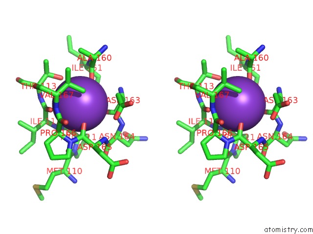
Stereo pair view
Mono view
Stereo pair view
A full contact list of Potassium with other atoms in the K binding
site number 1 of The Crystal Structure of Pyruvate Dehydrogenase E1 Bound to the Peripheral Subunit Binding Domain of E2 within 5.0Å range:
|
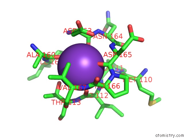
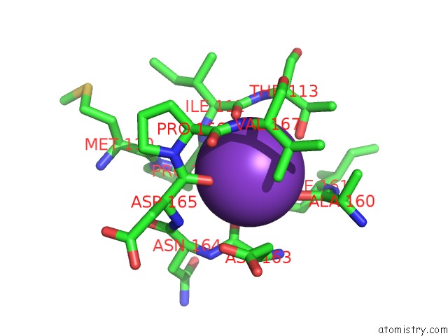
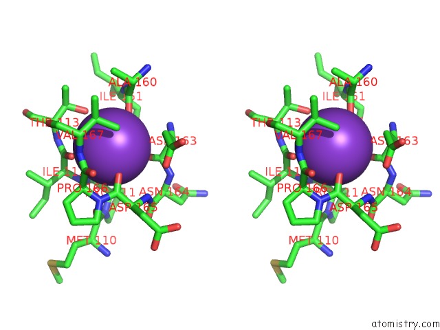
Potassium binding site 2 out of 4 in 1w85
Go back to
Potassium binding site 2 out
of 4 in the The Crystal Structure of Pyruvate Dehydrogenase E1 Bound to the Peripheral Subunit Binding Domain of E2
Mono view
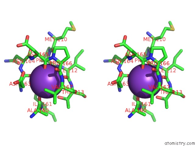
Stereo pair view
Mono view
Stereo pair view
A full contact list of Potassium with other atoms in the K binding
site number 2 of The Crystal Structure of Pyruvate Dehydrogenase E1 Bound to the Peripheral Subunit Binding Domain of E2 within 5.0Å range:
|
Potassium binding site 3 out of 4 in 1w85
Go back to
Potassium binding site 3 out
of 4 in the The Crystal Structure of Pyruvate Dehydrogenase E1 Bound to the Peripheral Subunit Binding Domain of E2
Mono view
Stereo pair view
Mono view
Stereo pair view
A full contact list of Potassium with other atoms in the K binding
site number 3 of The Crystal Structure of Pyruvate Dehydrogenase E1 Bound to the Peripheral Subunit Binding Domain of E2 within 5.0Å range:
|
Potassium binding site 4 out of 4 in 1w85
Go back to
Potassium binding site 4 out
of 4 in the The Crystal Structure of Pyruvate Dehydrogenase E1 Bound to the Peripheral Subunit Binding Domain of E2
Mono view
Stereo pair view
Mono view
Stereo pair view
A full contact list of Potassium with other atoms in the K binding
site number 4 of The Crystal Structure of Pyruvate Dehydrogenase E1 Bound to the Peripheral Subunit Binding Domain of E2 within 5.0Å range:
|
Reference:
R.A.Frank,
C.M.Titman,
J.V.Pratap,
B.F.Luisi,
R.N.Perham.
A Molecular Switch and Proton Wire Synchronize the Active Sites in Thiamine Enzymes. Science V. 306 872 2004.
ISSN: ESSN 1095-9203
PubMed: 15514159
DOI: 10.1126/SCIENCE.1101030
Page generated: Sat Aug 9 02:52:35 2025
ISSN: ESSN 1095-9203
PubMed: 15514159
DOI: 10.1126/SCIENCE.1101030
Last articles
K in 4TWKK in 4TS2
K in 4TOH
K in 4TOE
K in 4TOG
K in 4TOF
K in 4TOD
K in 4TOA
K in 4TO9
K in 4TM0