Potassium »
PDB 1u1g-1w29 »
1w22 »
Potassium in PDB 1w22: Crystal Structure of Inhibited Human HDAC8
Protein crystallography data
The structure of Crystal Structure of Inhibited Human HDAC8, PDB code: 1w22
was solved by
A.Vannini,
C.Volpari,
E.Caroli Casavola,
S.Di Marco,
with X-Ray Crystallography technique. A brief refinement statistics is given in the table below:
| Resolution Low / High (Å) | 50.0 / 2.5 |
| Space group | P 1 21 1 |
| Cell size a, b, c (Å), α, β, γ (°) | 51.746, 83.532, 94.653, 90.00, 97.48, 90.00 |
| R / Rfree (%) | n/a / n/a |
Other elements in 1w22:
The structure of Crystal Structure of Inhibited Human HDAC8 also contains other interesting chemical elements:
| Zinc | (Zn) | 2 atoms |
Potassium Binding Sites:
The binding sites of Potassium atom in the Crystal Structure of Inhibited Human HDAC8
(pdb code 1w22). This binding sites where shown within
5.0 Angstroms radius around Potassium atom.
In total 4 binding sites of Potassium where determined in the Crystal Structure of Inhibited Human HDAC8, PDB code: 1w22:
Jump to Potassium binding site number: 1; 2; 3; 4;
In total 4 binding sites of Potassium where determined in the Crystal Structure of Inhibited Human HDAC8, PDB code: 1w22:
Jump to Potassium binding site number: 1; 2; 3; 4;
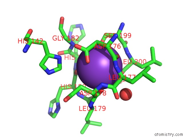
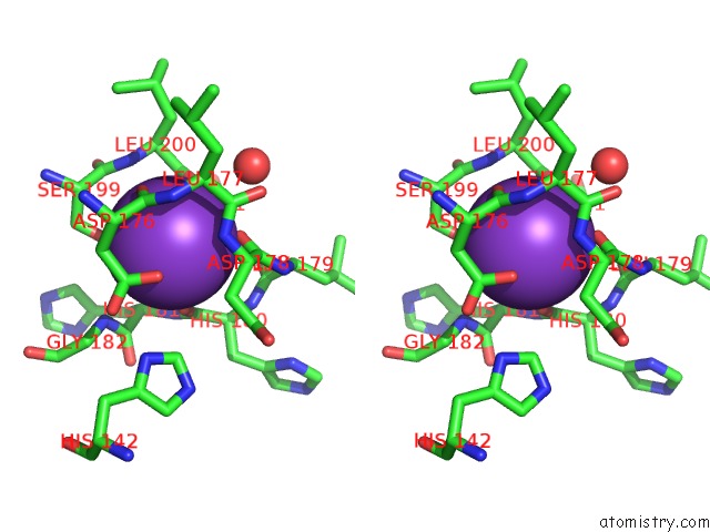
Potassium binding site 1 out of 4 in 1w22
Go back to
Potassium binding site 1 out
of 4 in the Crystal Structure of Inhibited Human HDAC8
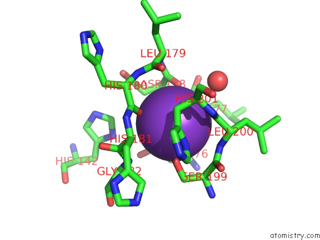
Mono view
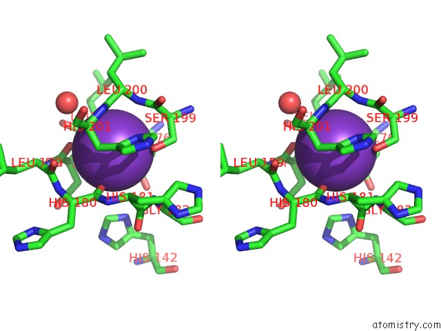
Stereo pair view
Mono view
Stereo pair view
A full contact list of Potassium with other atoms in the K binding
site number 1 of Crystal Structure of Inhibited Human HDAC8 within 5.0Å range:
|
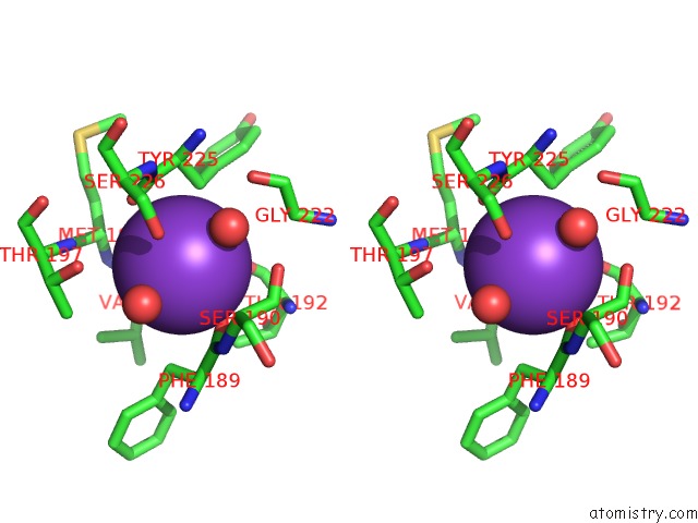
Potassium binding site 2 out of 4 in 1w22
Go back to
Potassium binding site 2 out
of 4 in the Crystal Structure of Inhibited Human HDAC8
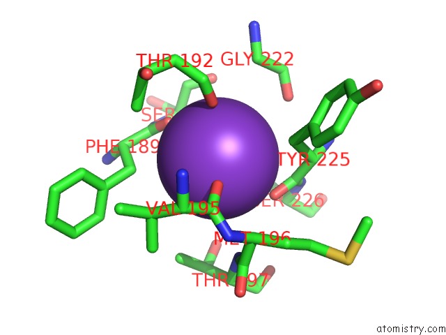
Mono view
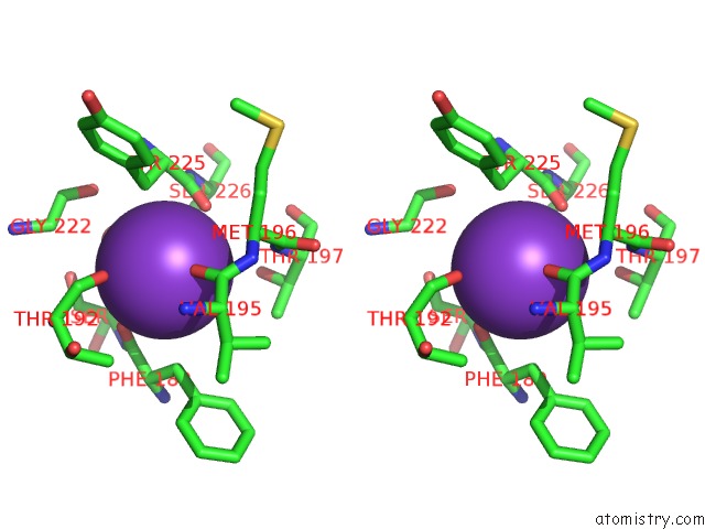
Stereo pair view
Mono view
Stereo pair view
A full contact list of Potassium with other atoms in the K binding
site number 2 of Crystal Structure of Inhibited Human HDAC8 within 5.0Å range:
|
Potassium binding site 3 out of 4 in 1w22
Go back to
Potassium binding site 3 out
of 4 in the Crystal Structure of Inhibited Human HDAC8
Mono view
Stereo pair view
Mono view
Stereo pair view
A full contact list of Potassium with other atoms in the K binding
site number 3 of Crystal Structure of Inhibited Human HDAC8 within 5.0Å range:
|
Potassium binding site 4 out of 4 in 1w22
Go back to
Potassium binding site 4 out
of 4 in the Crystal Structure of Inhibited Human HDAC8

Mono view
Stereo pair view
Mono view
Stereo pair view
A full contact list of Potassium with other atoms in the K binding
site number 4 of Crystal Structure of Inhibited Human HDAC8 within 5.0Å range:
|
Reference:
A.Vannini,
C.Volpari,
G.Filocamo,
E.Caroli Casavola,
M.Brunetti,
D.Renzoni,
P.Chakravarty,
C.Paolini,
R.De Francesco,
P.Gallinari,
C.Steinckuhler,
S.Di Marco.
Crystal Structure of A Eukaryotic Zn-Dependent Histone Deacetylase,Human HDAC8,Complexed with A Hydroxamic Acid Inhibitor Proc.Natl.Acad.Sci.Usa V. 101 15064 2004.
ISSN: ISSN 0027-8424
PubMed: 15477595
DOI: 10.1073/PNAS.0404603101
Page generated: Mon Aug 12 05:40:06 2024
ISSN: ISSN 0027-8424
PubMed: 15477595
DOI: 10.1073/PNAS.0404603101
Last articles
Zn in 9J0NZn in 9J0O
Zn in 9J0P
Zn in 9FJX
Zn in 9EKB
Zn in 9C0F
Zn in 9CAH
Zn in 9CH0
Zn in 9CH3
Zn in 9CH1