Potassium »
PDB 1u1g-1w29 »
1uc5 »
Potassium in PDB 1uc5: Structure of Diol Dehydratase Complexed with (R)-1,2- Propanediol
Enzymatic activity of Structure of Diol Dehydratase Complexed with (R)-1,2- Propanediol
All present enzymatic activity of Structure of Diol Dehydratase Complexed with (R)-1,2- Propanediol:
4.2.1.28;
4.2.1.28;
Protein crystallography data
The structure of Structure of Diol Dehydratase Complexed with (R)-1,2- Propanediol, PDB code: 1uc5
was solved by
N.Shibata,
Y.Nakanishi,
M.Fukuoka,
M.Yamanishi,
N.Yasuoka,
T.Toraya,
with X-Ray Crystallography technique. A brief refinement statistics is given in the table below:
| Resolution Low / High (Å) | 46.85 / 2.30 |
| Space group | P 21 21 21 |
| Cell size a, b, c (Å), α, β, γ (°) | 74.640, 122.270, 207.430, 90.00, 90.00, 90.00 |
| R / Rfree (%) | 20.8 / 25.5 |
Other elements in 1uc5:
The structure of Structure of Diol Dehydratase Complexed with (R)-1,2- Propanediol also contains other interesting chemical elements:
| Cobalt | (Co) | 2 atoms |
Potassium Binding Sites:
The binding sites of Potassium atom in the Structure of Diol Dehydratase Complexed with (R)-1,2- Propanediol
(pdb code 1uc5). This binding sites where shown within
5.0 Angstroms radius around Potassium atom.
In total 2 binding sites of Potassium where determined in the Structure of Diol Dehydratase Complexed with (R)-1,2- Propanediol, PDB code: 1uc5:
Jump to Potassium binding site number: 1; 2;
In total 2 binding sites of Potassium where determined in the Structure of Diol Dehydratase Complexed with (R)-1,2- Propanediol, PDB code: 1uc5:
Jump to Potassium binding site number: 1; 2;
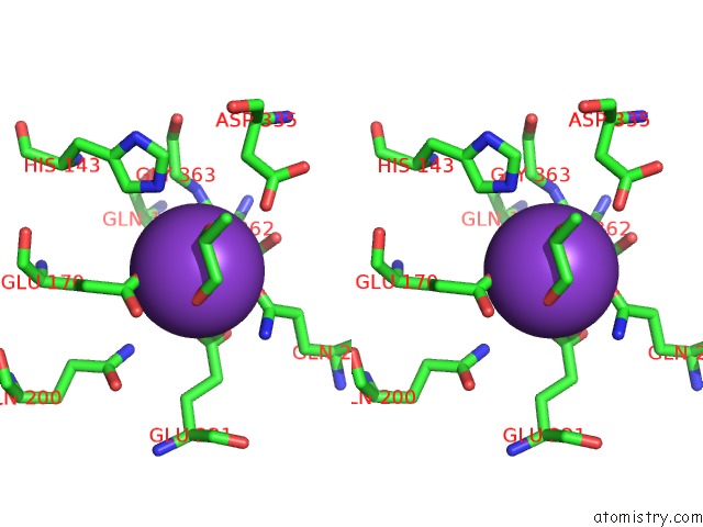
Potassium binding site 1 out of 2 in 1uc5
Go back to
Potassium binding site 1 out
of 2 in the Structure of Diol Dehydratase Complexed with (R)-1,2- Propanediol

Mono view
Stereo pair view
Mono view
Stereo pair view
A full contact list of Potassium with other atoms in the K binding
site number 1 of Structure of Diol Dehydratase Complexed with (R)-1,2- Propanediol within 5.0Å range:
|
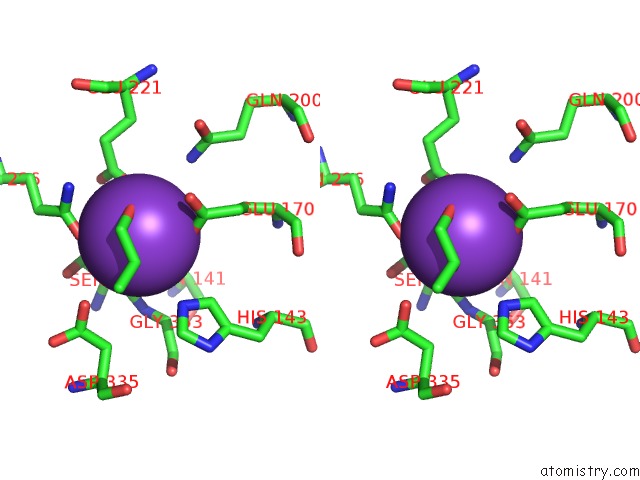
Potassium binding site 2 out of 2 in 1uc5
Go back to
Potassium binding site 2 out
of 2 in the Structure of Diol Dehydratase Complexed with (R)-1,2- Propanediol

Mono view
Stereo pair view
Mono view
Stereo pair view
A full contact list of Potassium with other atoms in the K binding
site number 2 of Structure of Diol Dehydratase Complexed with (R)-1,2- Propanediol within 5.0Å range:
|
Reference:
N.Shibata,
Y.Nakanishi,
M.Fukuoka,
M.Yamanishi,
N.Yasuoka,
T.Toraya.
Structural Rationalization For the Lack of Stereospecificity in Coenzyme B12-Dependent Diol Dehydratase J.Biol.Chem. V. 278 22717 2003.
ISSN: ISSN 0021-9258
PubMed: 12684496
DOI: 10.1074/JBC.M301513200
Page generated: Mon Aug 12 05:34:18 2024
ISSN: ISSN 0021-9258
PubMed: 12684496
DOI: 10.1074/JBC.M301513200
Last articles
Zn in 9MJ5Zn in 9HNW
Zn in 9G0L
Zn in 9FNE
Zn in 9DZN
Zn in 9E0I
Zn in 9D32
Zn in 9DAK
Zn in 8ZXC
Zn in 8ZUF