Potassium »
PDB 1s72-1u1f »
1u1d »
Potassium in PDB 1u1d: Structure of E. Coli Uridine Phosphorylase Complexed to 5- (Phenylthio)Acyclouridine (Ptau)
Enzymatic activity of Structure of E. Coli Uridine Phosphorylase Complexed to 5- (Phenylthio)Acyclouridine (Ptau)
All present enzymatic activity of Structure of E. Coli Uridine Phosphorylase Complexed to 5- (Phenylthio)Acyclouridine (Ptau):
2.4.2.3;
2.4.2.3;
Protein crystallography data
The structure of Structure of E. Coli Uridine Phosphorylase Complexed to 5- (Phenylthio)Acyclouridine (Ptau), PDB code: 1u1d
was solved by
W.Bu,
E.C.Settembre,
S.E.Ealick,
with X-Ray Crystallography technique. A brief refinement statistics is given in the table below:
| Resolution Low / High (Å) | 47.14 / 2.00 |
| Space group | P 21 21 21 |
| Cell size a, b, c (Å), α, β, γ (°) | 91.150, 125.746, 140.880, 90.00, 90.00, 90.00 |
| R / Rfree (%) | 20.9 / 22.6 |
Potassium Binding Sites:
The binding sites of Potassium atom in the Structure of E. Coli Uridine Phosphorylase Complexed to 5- (Phenylthio)Acyclouridine (Ptau)
(pdb code 1u1d). This binding sites where shown within
5.0 Angstroms radius around Potassium atom.
In total 3 binding sites of Potassium where determined in the Structure of E. Coli Uridine Phosphorylase Complexed to 5- (Phenylthio)Acyclouridine (Ptau), PDB code: 1u1d:
Jump to Potassium binding site number: 1; 2; 3;
In total 3 binding sites of Potassium where determined in the Structure of E. Coli Uridine Phosphorylase Complexed to 5- (Phenylthio)Acyclouridine (Ptau), PDB code: 1u1d:
Jump to Potassium binding site number: 1; 2; 3;
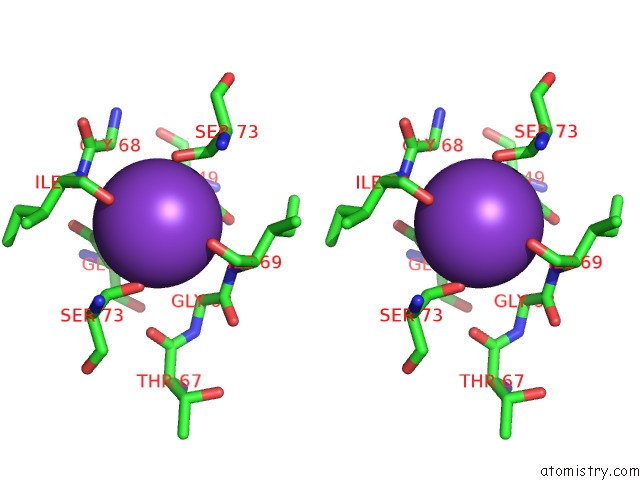
Potassium binding site 1 out of 3 in 1u1d
Go back to
Potassium binding site 1 out
of 3 in the Structure of E. Coli Uridine Phosphorylase Complexed to 5- (Phenylthio)Acyclouridine (Ptau)
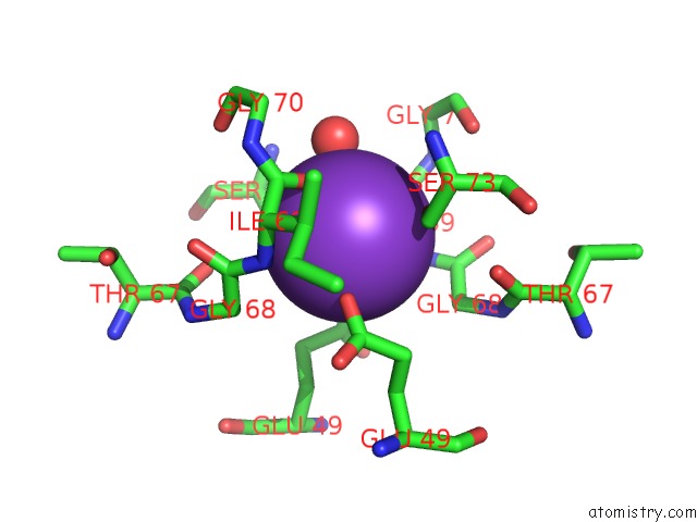
Mono view
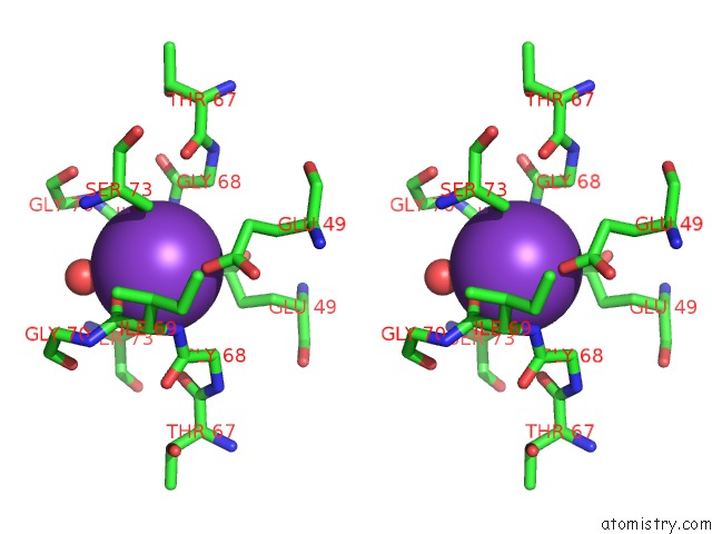
Stereo pair view
Mono view
Stereo pair view
A full contact list of Potassium with other atoms in the K binding
site number 1 of Structure of E. Coli Uridine Phosphorylase Complexed to 5- (Phenylthio)Acyclouridine (Ptau) within 5.0Å range:
|
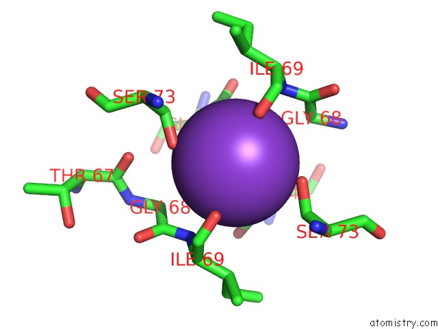
Potassium binding site 2 out of 3 in 1u1d
Go back to
Potassium binding site 2 out
of 3 in the Structure of E. Coli Uridine Phosphorylase Complexed to 5- (Phenylthio)Acyclouridine (Ptau)
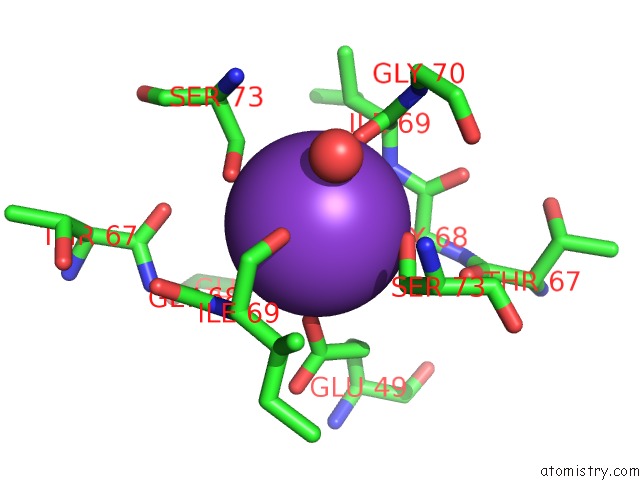
Mono view

Stereo pair view
Mono view
Stereo pair view
A full contact list of Potassium with other atoms in the K binding
site number 2 of Structure of E. Coli Uridine Phosphorylase Complexed to 5- (Phenylthio)Acyclouridine (Ptau) within 5.0Å range:
|
Potassium binding site 3 out of 3 in 1u1d
Go back to
Potassium binding site 3 out
of 3 in the Structure of E. Coli Uridine Phosphorylase Complexed to 5- (Phenylthio)Acyclouridine (Ptau)
Mono view
Stereo pair view
Mono view
Stereo pair view
A full contact list of Potassium with other atoms in the K binding
site number 3 of Structure of E. Coli Uridine Phosphorylase Complexed to 5- (Phenylthio)Acyclouridine (Ptau) within 5.0Å range:
|
Reference:
W.Bu,
E.C.Settembre,
M.H.El Kouni,
S.E.Ealick.
Structural Basis For Inhibition of Escherichia Coli Uridine Phosphorylase By 5-Substituted Acyclouridines. Acta Crystallogr.,Sect.D V. 61 863 2005.
ISSN: ISSN 0907-4449
PubMed: 15983408
DOI: 10.1107/S0907444905007882
Page generated: Mon Aug 12 05:32:35 2024
ISSN: ISSN 0907-4449
PubMed: 15983408
DOI: 10.1107/S0907444905007882
Last articles
Zn in 9J0NZn in 9J0O
Zn in 9J0P
Zn in 9FJX
Zn in 9EKB
Zn in 9C0F
Zn in 9CAH
Zn in 9CH0
Zn in 9CH3
Zn in 9CH1