Potassium »
PDB 1s72-1u1f »
1u12 »
Potassium in PDB 1u12: M. Loti Cyclic Nucleotide Binding Domain Mutant
Protein crystallography data
The structure of M. Loti Cyclic Nucleotide Binding Domain Mutant, PDB code: 1u12
was solved by
G.M.Clayton,
W.R.Silverman,
L.Heginbotham,
J.H.Morais-Cabral,
with X-Ray Crystallography technique. A brief refinement statistics is given in the table below:
| Resolution Low / High (Å) | 40.39 / 2.70 |
| Space group | P 41 21 2 |
| Cell size a, b, c (Å), α, β, γ (°) | 57.126, 57.126, 193.037, 90.00, 90.00, 90.00 |
| R / Rfree (%) | 25.3 / 28.1 |
Other elements in 1u12:
The structure of M. Loti Cyclic Nucleotide Binding Domain Mutant also contains other interesting chemical elements:
| Iodine | (I) | 31 atoms |
Potassium Binding Sites:
The binding sites of Potassium atom in the M. Loti Cyclic Nucleotide Binding Domain Mutant
(pdb code 1u12). This binding sites where shown within
5.0 Angstroms radius around Potassium atom.
In total 4 binding sites of Potassium where determined in the M. Loti Cyclic Nucleotide Binding Domain Mutant, PDB code: 1u12:
Jump to Potassium binding site number: 1; 2; 3; 4;
In total 4 binding sites of Potassium where determined in the M. Loti Cyclic Nucleotide Binding Domain Mutant, PDB code: 1u12:
Jump to Potassium binding site number: 1; 2; 3; 4;
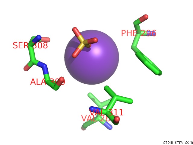
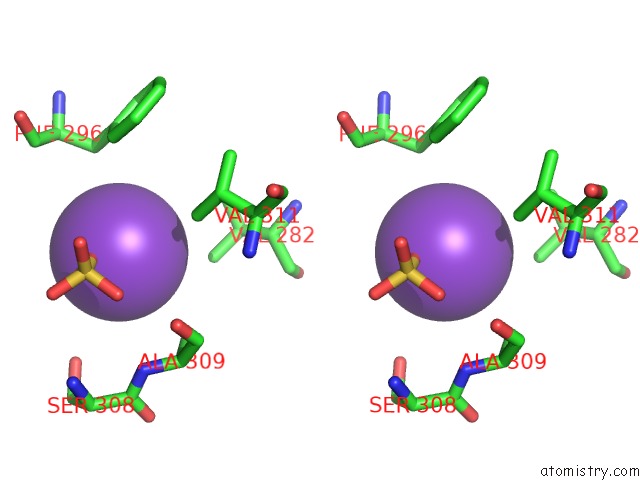
Potassium binding site 1 out of 4 in 1u12
Go back to
Potassium binding site 1 out
of 4 in the M. Loti Cyclic Nucleotide Binding Domain Mutant
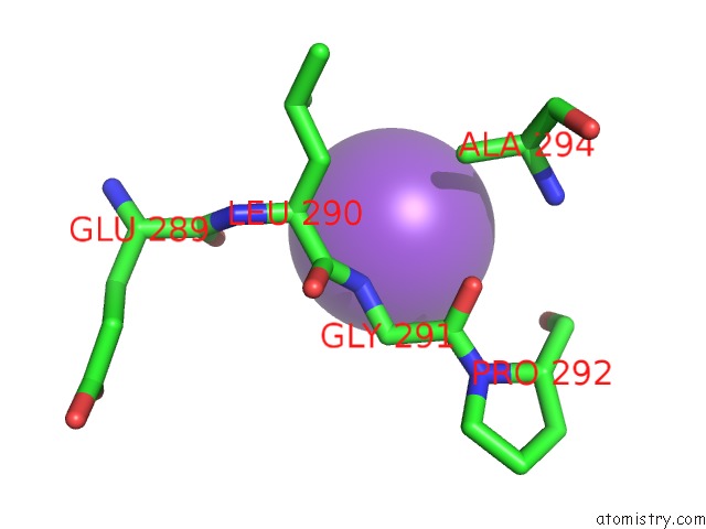
Mono view
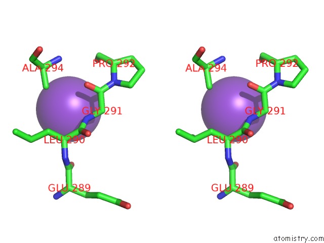
Stereo pair view
Mono view
Stereo pair view
A full contact list of Potassium with other atoms in the K binding
site number 1 of M. Loti Cyclic Nucleotide Binding Domain Mutant within 5.0Å range:
|
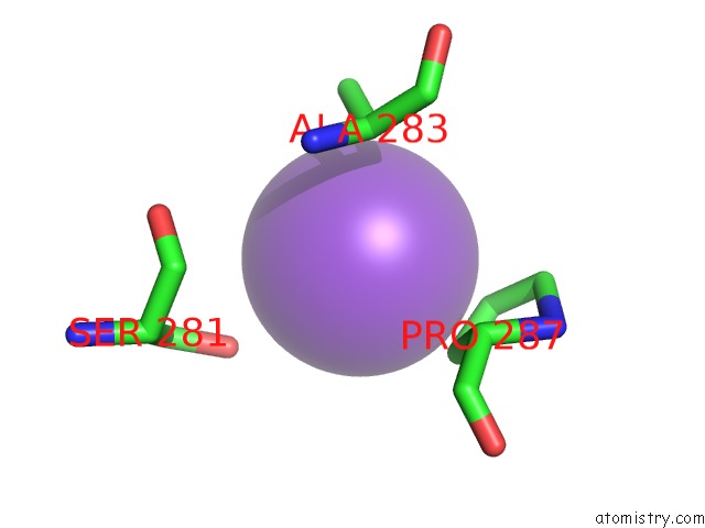
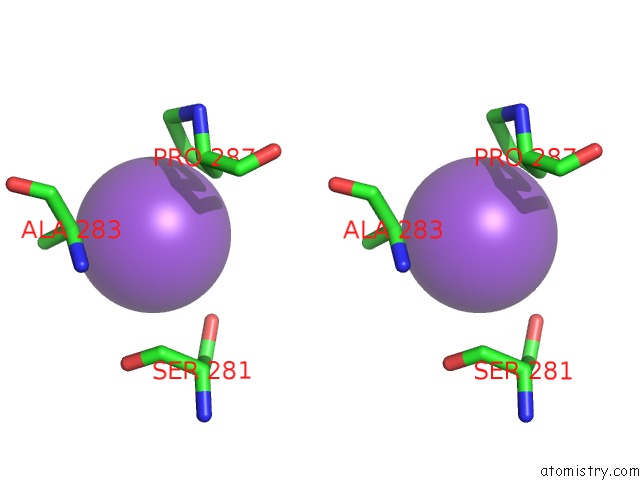
Potassium binding site 2 out of 4 in 1u12
Go back to
Potassium binding site 2 out
of 4 in the M. Loti Cyclic Nucleotide Binding Domain Mutant
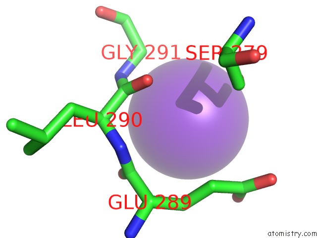
Mono view
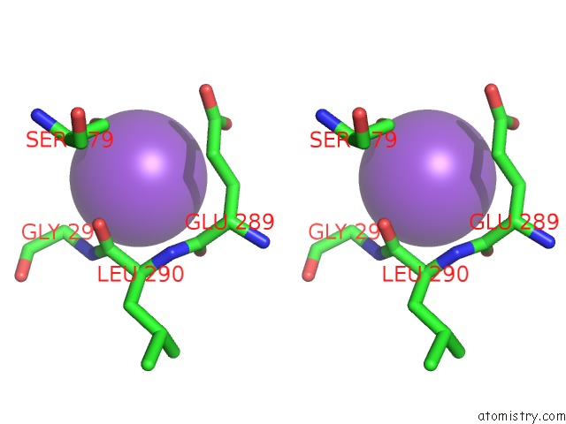
Stereo pair view
Mono view
Stereo pair view
A full contact list of Potassium with other atoms in the K binding
site number 2 of M. Loti Cyclic Nucleotide Binding Domain Mutant within 5.0Å range:
|
Potassium binding site 3 out of 4 in 1u12
Go back to
Potassium binding site 3 out
of 4 in the M. Loti Cyclic Nucleotide Binding Domain Mutant
Mono view
Stereo pair view
Mono view
Stereo pair view
A full contact list of Potassium with other atoms in the K binding
site number 3 of M. Loti Cyclic Nucleotide Binding Domain Mutant within 5.0Å range:
|
Potassium binding site 4 out of 4 in 1u12
Go back to
Potassium binding site 4 out
of 4 in the M. Loti Cyclic Nucleotide Binding Domain Mutant
Mono view
Stereo pair view
Mono view
Stereo pair view
A full contact list of Potassium with other atoms in the K binding
site number 4 of M. Loti Cyclic Nucleotide Binding Domain Mutant within 5.0Å range:
|
Reference:
G.M.Clayton,
W.R.Silverman,
L.Heginbotham,
J.H.Morais-Cabral.
Structural Basis of Ligand Activation in A Cyclic Nucleotide Regulated Potassium Channel Cell(Cambridge,Mass.) V. 119 615 2004.
ISSN: ISSN 0092-8674
PubMed: 15550244
DOI: 10.1016/J.CELL.2004.10.030
Page generated: Mon Aug 12 05:31:36 2024
ISSN: ISSN 0092-8674
PubMed: 15550244
DOI: 10.1016/J.CELL.2004.10.030
Last articles
Zn in 9J0NZn in 9J0O
Zn in 9J0P
Zn in 9FJX
Zn in 9EKB
Zn in 9C0F
Zn in 9CAH
Zn in 9CH0
Zn in 9CH3
Zn in 9CH1