Potassium »
PDB 1s72-1u1f »
1t9d »
Potassium in PDB 1t9d: Crystal Structure of Yeast Acetohydroxyacid Synthase in Complex with A Sulfonylurea Herbicide, Metsulfuron Methyl
Enzymatic activity of Crystal Structure of Yeast Acetohydroxyacid Synthase in Complex with A Sulfonylurea Herbicide, Metsulfuron Methyl
All present enzymatic activity of Crystal Structure of Yeast Acetohydroxyacid Synthase in Complex with A Sulfonylurea Herbicide, Metsulfuron Methyl:
2.2.1.6;
2.2.1.6;
Protein crystallography data
The structure of Crystal Structure of Yeast Acetohydroxyacid Synthase in Complex with A Sulfonylurea Herbicide, Metsulfuron Methyl, PDB code: 1t9d
was solved by
J.A.Mccourt,
S.S.Pang,
L.W.Guddat,
R.G.Duggleby,
with X-Ray Crystallography technique. A brief refinement statistics is given in the table below:
| Resolution Low / High (Å) | 50.00 / 2.30 |
| Space group | I 4 2 2 |
| Cell size a, b, c (Å), α, β, γ (°) | 218.347, 218.347, 361.530, 90.00, 90.00, 90.00 |
| R / Rfree (%) | 16.4 / 19.5 |
Other elements in 1t9d:
The structure of Crystal Structure of Yeast Acetohydroxyacid Synthase in Complex with A Sulfonylurea Herbicide, Metsulfuron Methyl also contains other interesting chemical elements:
| Magnesium | (Mg) | 4 atoms |
Potassium Binding Sites:
The binding sites of Potassium atom in the Crystal Structure of Yeast Acetohydroxyacid Synthase in Complex with A Sulfonylurea Herbicide, Metsulfuron Methyl
(pdb code 1t9d). This binding sites where shown within
5.0 Angstroms radius around Potassium atom.
In total 4 binding sites of Potassium where determined in the Crystal Structure of Yeast Acetohydroxyacid Synthase in Complex with A Sulfonylurea Herbicide, Metsulfuron Methyl, PDB code: 1t9d:
Jump to Potassium binding site number: 1; 2; 3; 4;
In total 4 binding sites of Potassium where determined in the Crystal Structure of Yeast Acetohydroxyacid Synthase in Complex with A Sulfonylurea Herbicide, Metsulfuron Methyl, PDB code: 1t9d:
Jump to Potassium binding site number: 1; 2; 3; 4;
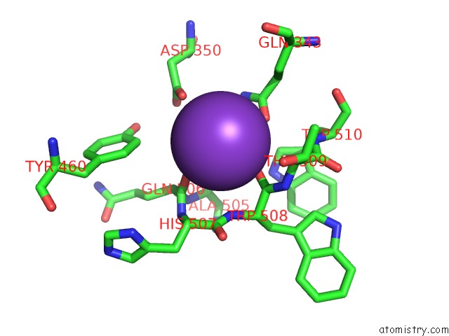
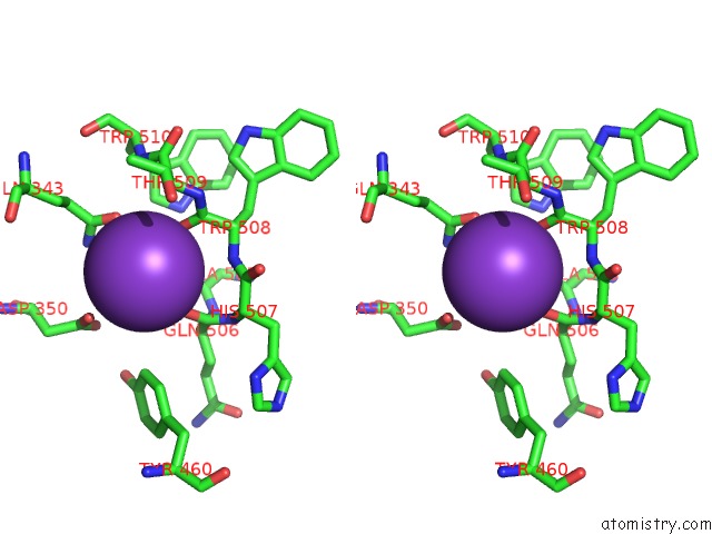
Potassium binding site 1 out of 4 in 1t9d
Go back to
Potassium binding site 1 out
of 4 in the Crystal Structure of Yeast Acetohydroxyacid Synthase in Complex with A Sulfonylurea Herbicide, Metsulfuron Methyl
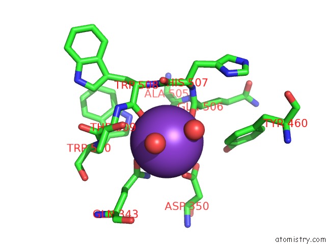
Mono view
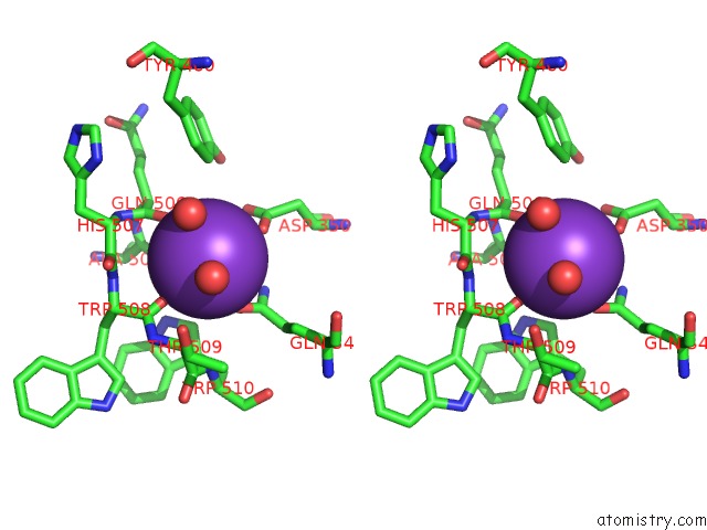
Stereo pair view
Mono view
Stereo pair view
A full contact list of Potassium with other atoms in the K binding
site number 1 of Crystal Structure of Yeast Acetohydroxyacid Synthase in Complex with A Sulfonylurea Herbicide, Metsulfuron Methyl within 5.0Å range:
|
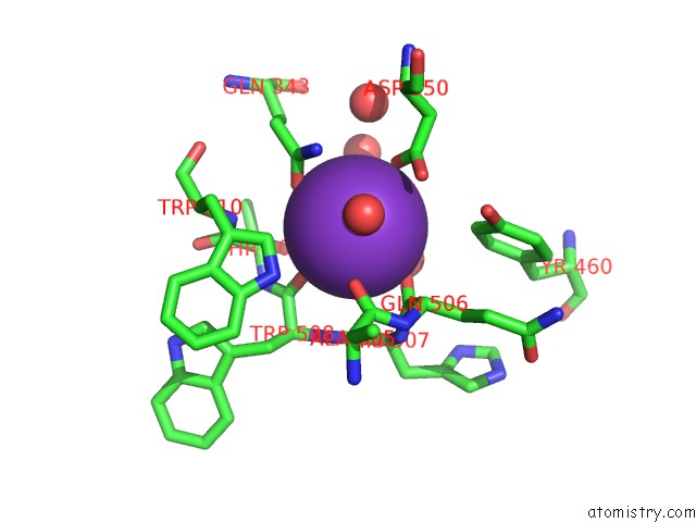
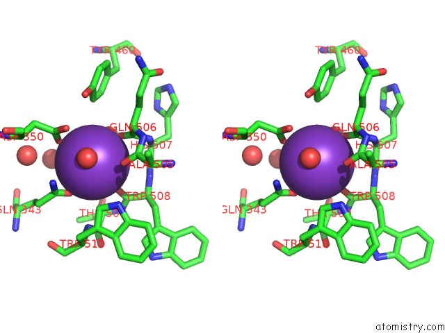
Potassium binding site 2 out of 4 in 1t9d
Go back to
Potassium binding site 2 out
of 4 in the Crystal Structure of Yeast Acetohydroxyacid Synthase in Complex with A Sulfonylurea Herbicide, Metsulfuron Methyl
Mono view
Stereo pair view
Mono view
Stereo pair view
A full contact list of Potassium with other atoms in the K binding
site number 2 of Crystal Structure of Yeast Acetohydroxyacid Synthase in Complex with A Sulfonylurea Herbicide, Metsulfuron Methyl within 5.0Å range:
|
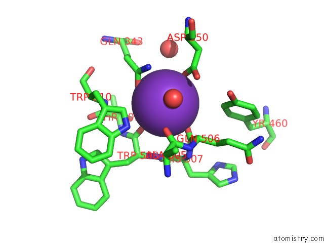
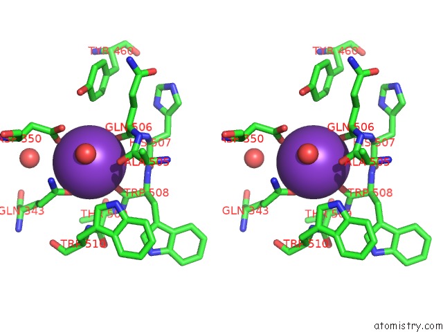
Potassium binding site 3 out of 4 in 1t9d
Go back to
Potassium binding site 3 out
of 4 in the Crystal Structure of Yeast Acetohydroxyacid Synthase in Complex with A Sulfonylurea Herbicide, Metsulfuron Methyl
Mono view
Stereo pair view
Mono view
Stereo pair view
A full contact list of Potassium with other atoms in the K binding
site number 3 of Crystal Structure of Yeast Acetohydroxyacid Synthase in Complex with A Sulfonylurea Herbicide, Metsulfuron Methyl within 5.0Å range:
|
Potassium binding site 4 out of 4 in 1t9d
Go back to
Potassium binding site 4 out
of 4 in the Crystal Structure of Yeast Acetohydroxyacid Synthase in Complex with A Sulfonylurea Herbicide, Metsulfuron Methyl
Mono view
Stereo pair view
Mono view
Stereo pair view
A full contact list of Potassium with other atoms in the K binding
site number 4 of Crystal Structure of Yeast Acetohydroxyacid Synthase in Complex with A Sulfonylurea Herbicide, Metsulfuron Methyl within 5.0Å range:
|
Reference:
J.A.Mccourt,
S.S.Pang,
L.W.Guddat,
R.G.Duggleby.
Elucidating the Specificity of Binding of Sulfonylurea Herbicides to Acetohydroxyacid Synthase. Biochemistry V. 44 2330 2005.
ISSN: ISSN 0006-2960
PubMed: 15709745
DOI: 10.1021/BI047980A
Page generated: Mon Aug 12 05:28:36 2024
ISSN: ISSN 0006-2960
PubMed: 15709745
DOI: 10.1021/BI047980A
Last articles
Zn in 9J0NZn in 9J0O
Zn in 9J0P
Zn in 9FJX
Zn in 9EKB
Zn in 9C0F
Zn in 9CAH
Zn in 9CH0
Zn in 9CH3
Zn in 9CH1