Potassium »
PDB 1pr9-1s61 »
1rxy »
Potassium in PDB 1rxy: E. Coli Uridine Phosphorylase: Type-B Native
Enzymatic activity of E. Coli Uridine Phosphorylase: Type-B Native
All present enzymatic activity of E. Coli Uridine Phosphorylase: Type-B Native:
2.4.2.3;
2.4.2.3;
Protein crystallography data
The structure of E. Coli Uridine Phosphorylase: Type-B Native, PDB code: 1rxy
was solved by
T.T.Caradoc-Davies,
S.M.Cutfield,
I.L.Lamont,
J.F.Cutfield,
with X-Ray Crystallography technique. A brief refinement statistics is given in the table below:
| Resolution Low / High (Å) | 16.10 / 1.70 |
| Space group | H 3 |
| Cell size a, b, c (Å), α, β, γ (°) | 152.928, 152.928, 50.864, 90.00, 90.00, 120.00 |
| R / Rfree (%) | 12.8 / 16.4 |
Potassium Binding Sites:
The binding sites of Potassium atom in the E. Coli Uridine Phosphorylase: Type-B Native
(pdb code 1rxy). This binding sites where shown within
5.0 Angstroms radius around Potassium atom.
In total only one binding site of Potassium was determined in the E. Coli Uridine Phosphorylase: Type-B Native, PDB code: 1rxy:
In total only one binding site of Potassium was determined in the E. Coli Uridine Phosphorylase: Type-B Native, PDB code: 1rxy:
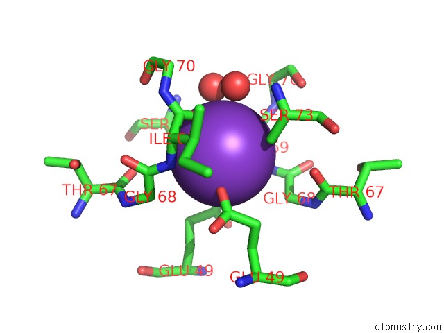
Potassium binding site 1 out of 1 in 1rxy
Go back to
Potassium binding site 1 out
of 1 in the E. Coli Uridine Phosphorylase: Type-B Native
Mono view

Stereo pair view
Mono view
Stereo pair view
A full contact list of Potassium with other atoms in the K binding
site number 1 of E. Coli Uridine Phosphorylase: Type-B Native within 5.0Å range:
|
Reference:
T.T.Caradoc-Davies,
S.M.Cutfield,
I.L.Lamont,
J.F.Cutfield.
Crystal Structures of Escherichia Coli Uridine Phosphorylase in Two Native and Three Complexed Forms Reveal Basis of Substrate Specificity, Induced Conformational Changes and Influence of Potassium J.Mol.Biol. V. 337 337 2004.
ISSN: ISSN 0022-2836
PubMed: 15003451
DOI: 10.1016/J.JMB.2004.01.039
Page generated: Mon Aug 12 05:18:48 2024
ISSN: ISSN 0022-2836
PubMed: 15003451
DOI: 10.1016/J.JMB.2004.01.039
Last articles
Zn in 9J0NZn in 9J0O
Zn in 9J0P
Zn in 9FJX
Zn in 9EKB
Zn in 9C0F
Zn in 9CAH
Zn in 9CH0
Zn in 9CH3
Zn in 9CH1