Potassium »
PDB 1pr9-1s61 »
1rrv »
Potassium in PDB 1rrv: X-Ray Crystal Structure of Tdp-Vancosaminyltransferase Gtfd As A Complex with Tdp and the Natural Substrate, Desvancosaminyl Vancomycin.
Protein crystallography data
The structure of X-Ray Crystal Structure of Tdp-Vancosaminyltransferase Gtfd As A Complex with Tdp and the Natural Substrate, Desvancosaminyl Vancomycin., PDB code: 1rrv
was solved by
A.M.Mulichak,
W.Lu,
H.C.Losey,
C.T.Walsh,
R.M.Garavito,
with X-Ray Crystallography technique. A brief refinement statistics is given in the table below:
| Resolution Low / High (Å) | 30.00 / 2.00 |
| Space group | P 1 21 1 |
| Cell size a, b, c (Å), α, β, γ (°) | 50.670, 64.120, 144.080, 90.00, 91.73, 90.00 |
| R / Rfree (%) | 21 / 25.2 |
Other elements in 1rrv:
The structure of X-Ray Crystal Structure of Tdp-Vancosaminyltransferase Gtfd As A Complex with Tdp and the Natural Substrate, Desvancosaminyl Vancomycin. also contains other interesting chemical elements:
| Chlorine | (Cl) | 4 atoms |
Potassium Binding Sites:
The binding sites of Potassium atom in the X-Ray Crystal Structure of Tdp-Vancosaminyltransferase Gtfd As A Complex with Tdp and the Natural Substrate, Desvancosaminyl Vancomycin.
(pdb code 1rrv). This binding sites where shown within
5.0 Angstroms radius around Potassium atom.
In total 2 binding sites of Potassium where determined in the X-Ray Crystal Structure of Tdp-Vancosaminyltransferase Gtfd As A Complex with Tdp and the Natural Substrate, Desvancosaminyl Vancomycin., PDB code: 1rrv:
Jump to Potassium binding site number: 1; 2;
In total 2 binding sites of Potassium where determined in the X-Ray Crystal Structure of Tdp-Vancosaminyltransferase Gtfd As A Complex with Tdp and the Natural Substrate, Desvancosaminyl Vancomycin., PDB code: 1rrv:
Jump to Potassium binding site number: 1; 2;
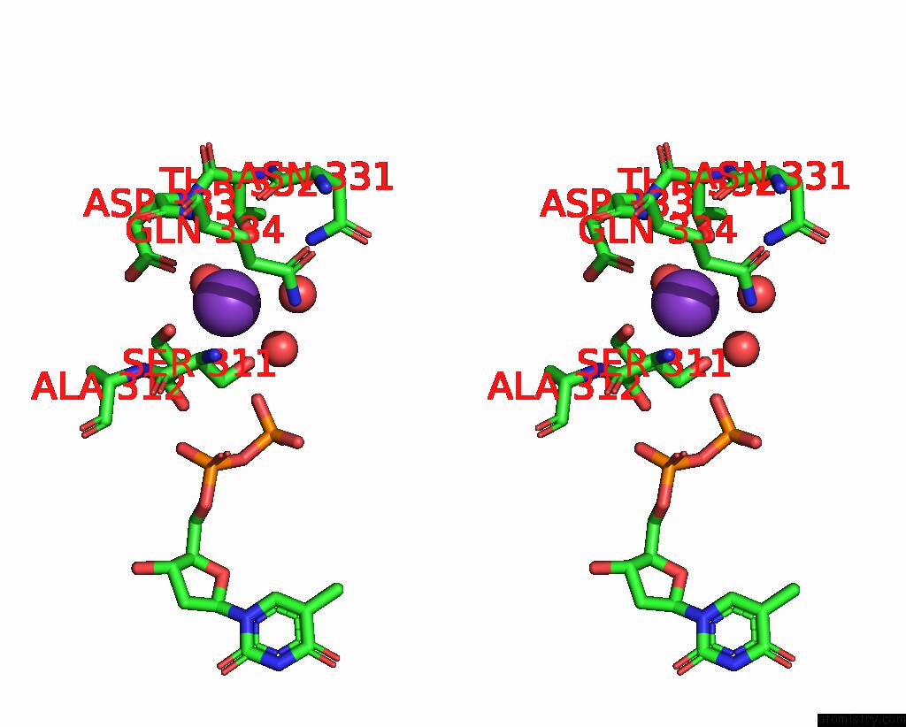
Potassium binding site 1 out of 2 in 1rrv
Go back to
Potassium binding site 1 out
of 2 in the X-Ray Crystal Structure of Tdp-Vancosaminyltransferase Gtfd As A Complex with Tdp and the Natural Substrate, Desvancosaminyl Vancomycin.

Mono view

Stereo pair view
Mono view
Stereo pair view
A full contact list of Potassium with other atoms in the K binding
site number 1 of X-Ray Crystal Structure of Tdp-Vancosaminyltransferase Gtfd As A Complex with Tdp and the Natural Substrate, Desvancosaminyl Vancomycin. within 5.0Å range:
|
Potassium binding site 2 out of 2 in 1rrv
Go back to
Potassium binding site 2 out
of 2 in the X-Ray Crystal Structure of Tdp-Vancosaminyltransferase Gtfd As A Complex with Tdp and the Natural Substrate, Desvancosaminyl Vancomycin.

Mono view
Stereo pair view
Mono view
Stereo pair view
A full contact list of Potassium with other atoms in the K binding
site number 2 of X-Ray Crystal Structure of Tdp-Vancosaminyltransferase Gtfd As A Complex with Tdp and the Natural Substrate, Desvancosaminyl Vancomycin. within 5.0Å range:
|
Reference:
A.M.Mulichak,
W.Lu,
H.C.Losey,
C.T.Walsh,
R.M.Garavito.
Crystal Structure of Vancosaminyltransferase Gtfd From the Vancomycin Biosynthetic Pathway: Interactions with Acceptor and Nucleotide Ligands Biochemistry V. 43 5170 2004.
ISSN: ISSN 0006-2960
PubMed: 15122882
DOI: 10.1021/BI036130C
Page generated: Mon Aug 12 05:17:57 2024
ISSN: ISSN 0006-2960
PubMed: 15122882
DOI: 10.1021/BI036130C
Last articles
Zn in 9J0NZn in 9J0O
Zn in 9J0P
Zn in 9FJX
Zn in 9EKB
Zn in 9C0F
Zn in 9CAH
Zn in 9CH0
Zn in 9CH3
Zn in 9CH1