Potassium »
PDB 1pr9-1s61 »
1rft »
Potassium in PDB 1rft: Crystal Structure of Pyridoxal Kinase Complexed with Amp- Pcp and Pyridoxamine
Enzymatic activity of Crystal Structure of Pyridoxal Kinase Complexed with Amp- Pcp and Pyridoxamine
All present enzymatic activity of Crystal Structure of Pyridoxal Kinase Complexed with Amp- Pcp and Pyridoxamine:
2.7.1.35;
2.7.1.35;
Protein crystallography data
The structure of Crystal Structure of Pyridoxal Kinase Complexed with Amp- Pcp and Pyridoxamine, PDB code: 1rft
was solved by
D.-C.Liang,
T.Jiang,
M.-H.Li,
with X-Ray Crystallography technique. A brief refinement statistics is given in the table below:
| Resolution Low / High (Å) | 20.00 / 2.80 |
| Space group | P 31 2 1 |
| Cell size a, b, c (Å), α, β, γ (°) | 103.687, 103.687, 58.604, 90.00, 90.00, 120.00 |
| R / Rfree (%) | 21.4 / 26.8 |
Other elements in 1rft:
The structure of Crystal Structure of Pyridoxal Kinase Complexed with Amp- Pcp and Pyridoxamine also contains other interesting chemical elements:
| Zinc | (Zn) | 1 atom |
Potassium Binding Sites:
The binding sites of Potassium atom in the Crystal Structure of Pyridoxal Kinase Complexed with Amp- Pcp and Pyridoxamine
(pdb code 1rft). This binding sites where shown within
5.0 Angstroms radius around Potassium atom.
In total only one binding site of Potassium was determined in the Crystal Structure of Pyridoxal Kinase Complexed with Amp- Pcp and Pyridoxamine, PDB code: 1rft:
In total only one binding site of Potassium was determined in the Crystal Structure of Pyridoxal Kinase Complexed with Amp- Pcp and Pyridoxamine, PDB code: 1rft:
Potassium binding site 1 out of 1 in 1rft
Go back to
Potassium binding site 1 out
of 1 in the Crystal Structure of Pyridoxal Kinase Complexed with Amp- Pcp and Pyridoxamine
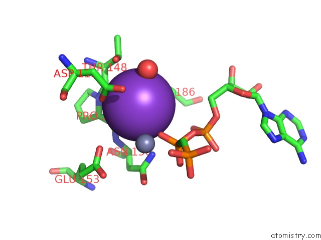
Mono view
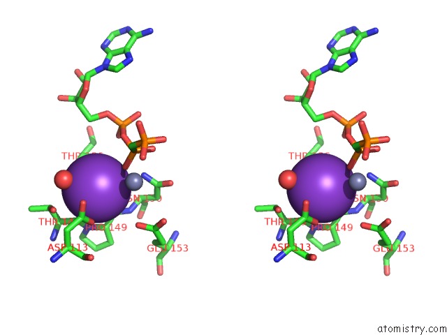
Stereo pair view
Mono view
Stereo pair view
A full contact list of Potassium with other atoms in the K binding
site number 1 of Crystal Structure of Pyridoxal Kinase Complexed with Amp- Pcp and Pyridoxamine within 5.0Å range:
|
Reference:
M.-H.Li,
F.Kwok,
W.-R.Chang,
S.-Q.Liu,
S.C.L.Lo,
J.-P.Zhang,
T.Jiang,
D.-C.Liang.
Conformational Changes in the Reaction of Pyridoxal Kinase J.Biol.Chem. V. 279 17459 2004.
ISSN: ISSN 0021-9258
PubMed: 14722069
DOI: 10.1074/JBC.M312380200
Page generated: Mon Aug 12 05:17:31 2024
ISSN: ISSN 0021-9258
PubMed: 14722069
DOI: 10.1074/JBC.M312380200
Last articles
Zn in 9MJ5Zn in 9HNW
Zn in 9G0L
Zn in 9FNE
Zn in 9DZN
Zn in 9E0I
Zn in 9D32
Zn in 9DAK
Zn in 8ZXC
Zn in 8ZUF