Potassium »
PDB 1pr9-1s61 »
1r64 »
Potassium in PDB 1r64: The 2.2 A Crystal Structure of KEX2 Protease in Complex with Ac-Arg- Glu-Lys-Boroarg Peptidyl Boronic Acid Inhibitor
Enzymatic activity of The 2.2 A Crystal Structure of KEX2 Protease in Complex with Ac-Arg- Glu-Lys-Boroarg Peptidyl Boronic Acid Inhibitor
All present enzymatic activity of The 2.2 A Crystal Structure of KEX2 Protease in Complex with Ac-Arg- Glu-Lys-Boroarg Peptidyl Boronic Acid Inhibitor:
3.4.21.61;
3.4.21.61;
Protein crystallography data
The structure of The 2.2 A Crystal Structure of KEX2 Protease in Complex with Ac-Arg- Glu-Lys-Boroarg Peptidyl Boronic Acid Inhibitor, PDB code: 1r64
was solved by
T.Holyoak,
C.A.Kettner,
G.A.Petsko,
R.S.Fuller,
D.Ringe,
with X-Ray Crystallography technique. A brief refinement statistics is given in the table below:
| Resolution Low / High (Å) | 50.00 / 2.20 |
| Space group | P 65 2 2 |
| Cell size a, b, c (Å), α, β, γ (°) | 113.541, 113.541, 364.971, 90.00, 90.00, 120.00 |
| R / Rfree (%) | 19.7 / 23.4 |
Other elements in 1r64:
The structure of The 2.2 A Crystal Structure of KEX2 Protease in Complex with Ac-Arg- Glu-Lys-Boroarg Peptidyl Boronic Acid Inhibitor also contains other interesting chemical elements:
| Calcium | (Ca) | 6 atoms |
Potassium Binding Sites:
The binding sites of Potassium atom in the The 2.2 A Crystal Structure of KEX2 Protease in Complex with Ac-Arg- Glu-Lys-Boroarg Peptidyl Boronic Acid Inhibitor
(pdb code 1r64). This binding sites where shown within
5.0 Angstroms radius around Potassium atom.
In total 6 binding sites of Potassium where determined in the The 2.2 A Crystal Structure of KEX2 Protease in Complex with Ac-Arg- Glu-Lys-Boroarg Peptidyl Boronic Acid Inhibitor, PDB code: 1r64:
Jump to Potassium binding site number: 1; 2; 3; 4; 5; 6;
In total 6 binding sites of Potassium where determined in the The 2.2 A Crystal Structure of KEX2 Protease in Complex with Ac-Arg- Glu-Lys-Boroarg Peptidyl Boronic Acid Inhibitor, PDB code: 1r64:
Jump to Potassium binding site number: 1; 2; 3; 4; 5; 6;
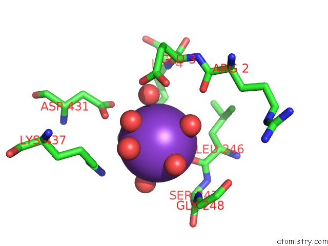
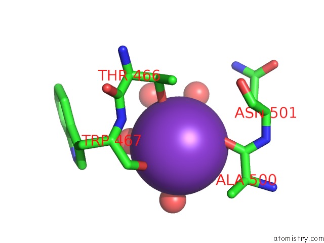
Potassium binding site 1 out of 6 in 1r64
Go back to
Potassium binding site 1 out
of 6 in the The 2.2 A Crystal Structure of KEX2 Protease in Complex with Ac-Arg- Glu-Lys-Boroarg Peptidyl Boronic Acid Inhibitor
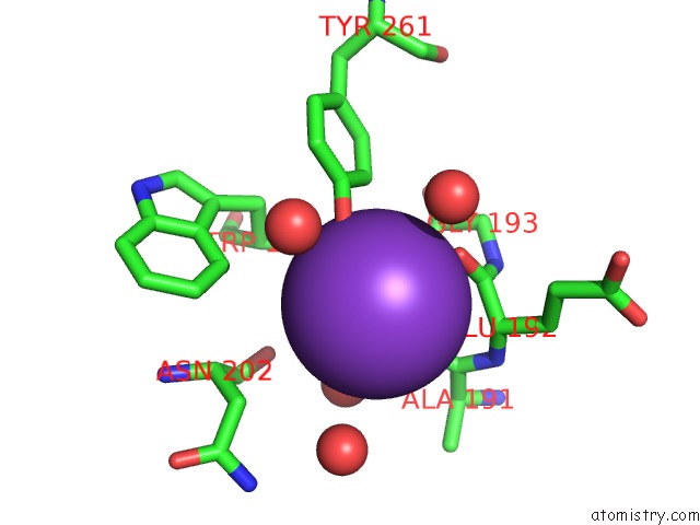
Mono view
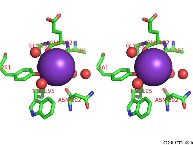
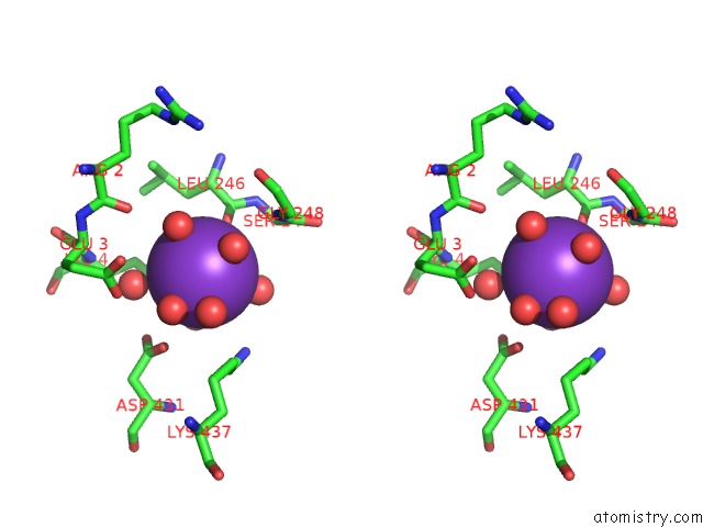
Stereo pair view
Mono view
Stereo pair view
A full contact list of Potassium with other atoms in the K binding
site number 1 of The 2.2 A Crystal Structure of KEX2 Protease in Complex with Ac-Arg- Glu-Lys-Boroarg Peptidyl Boronic Acid Inhibitor within 5.0Å range:
|
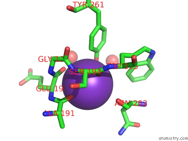
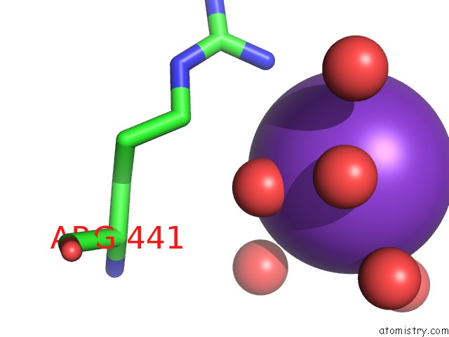
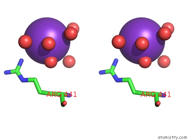
Potassium binding site 2 out of 6 in 1r64
Go back to
Potassium binding site 2 out
of 6 in the The 2.2 A Crystal Structure of KEX2 Protease in Complex with Ac-Arg- Glu-Lys-Boroarg Peptidyl Boronic Acid Inhibitor
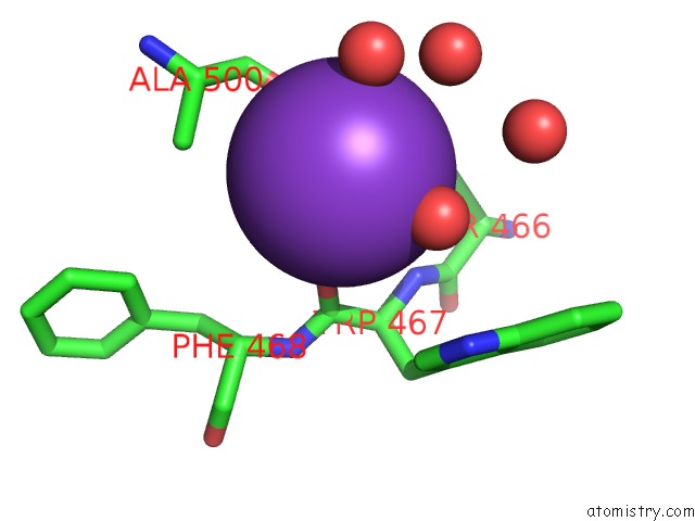
Mono view
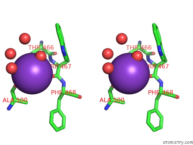
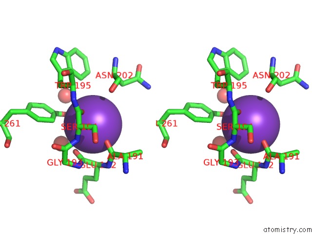
Stereo pair view
Mono view
Stereo pair view
A full contact list of Potassium with other atoms in the K binding
site number 2 of The 2.2 A Crystal Structure of KEX2 Protease in Complex with Ac-Arg- Glu-Lys-Boroarg Peptidyl Boronic Acid Inhibitor within 5.0Å range:
|
Potassium binding site 3 out of 6 in 1r64
Go back to
Potassium binding site 3 out
of 6 in the The 2.2 A Crystal Structure of KEX2 Protease in Complex with Ac-Arg- Glu-Lys-Boroarg Peptidyl Boronic Acid Inhibitor
Mono view
Stereo pair view
Mono view
Stereo pair view
A full contact list of Potassium with other atoms in the K binding
site number 3 of The 2.2 A Crystal Structure of KEX2 Protease in Complex with Ac-Arg- Glu-Lys-Boroarg Peptidyl Boronic Acid Inhibitor within 5.0Å range:
|
Potassium binding site 4 out of 6 in 1r64
Go back to
Potassium binding site 4 out
of 6 in the The 2.2 A Crystal Structure of KEX2 Protease in Complex with Ac-Arg- Glu-Lys-Boroarg Peptidyl Boronic Acid Inhibitor
Mono view
Stereo pair view
Mono view
Stereo pair view
A full contact list of Potassium with other atoms in the K binding
site number 4 of The 2.2 A Crystal Structure of KEX2 Protease in Complex with Ac-Arg- Glu-Lys-Boroarg Peptidyl Boronic Acid Inhibitor within 5.0Å range:
|
Potassium binding site 5 out of 6 in 1r64
Go back to
Potassium binding site 5 out
of 6 in the The 2.2 A Crystal Structure of KEX2 Protease in Complex with Ac-Arg- Glu-Lys-Boroarg Peptidyl Boronic Acid Inhibitor
Mono view

Stereo pair view
Mono view
Stereo pair view
A full contact list of Potassium with other atoms in the K binding
site number 5 of The 2.2 A Crystal Structure of KEX2 Protease in Complex with Ac-Arg- Glu-Lys-Boroarg Peptidyl Boronic Acid Inhibitor within 5.0Å range:
|
Potassium binding site 6 out of 6 in 1r64
Go back to
Potassium binding site 6 out
of 6 in the The 2.2 A Crystal Structure of KEX2 Protease in Complex with Ac-Arg- Glu-Lys-Boroarg Peptidyl Boronic Acid Inhibitor
Mono view
Stereo pair view
Mono view
Stereo pair view
A full contact list of Potassium with other atoms in the K binding
site number 6 of The 2.2 A Crystal Structure of KEX2 Protease in Complex with Ac-Arg- Glu-Lys-Boroarg Peptidyl Boronic Acid Inhibitor within 5.0Å range:
|
Reference:
T.Holyoak,
C.A.Kettner,
G.A.Petsko,
R.S.Fuller,
D.Ringe.
Structural Basis For Differences in Substrate Selectivity in KEX2 and Furin Protein Convertases Biochemistry V. 43 2412 2004.
ISSN: ISSN 0006-2960
PubMed: 14992578
DOI: 10.1021/BI035849H
Page generated: Mon Aug 12 05:16:43 2024
ISSN: ISSN 0006-2960
PubMed: 14992578
DOI: 10.1021/BI035849H
Last articles
Zn in 9J0NZn in 9J0O
Zn in 9J0P
Zn in 9FJX
Zn in 9EKB
Zn in 9C0F
Zn in 9CAH
Zn in 9CH0
Zn in 9CH3
Zn in 9CH1