Potassium »
PDB 1o76-1pqo »
1pkx »
Potassium in PDB 1pkx: Crystal Structure of Human Atic in Complex with Xmp
Enzymatic activity of Crystal Structure of Human Atic in Complex with Xmp
All present enzymatic activity of Crystal Structure of Human Atic in Complex with Xmp:
2.1.2.3; 3.5.4.10;
2.1.2.3; 3.5.4.10;
Protein crystallography data
The structure of Crystal Structure of Human Atic in Complex with Xmp, PDB code: 1pkx
was solved by
D.W.Wolan,
C.G.Cheong,
S.E.Greasley,
I.A.Wilson,
with X-Ray Crystallography technique. A brief refinement statistics is given in the table below:
| Resolution Low / High (Å) | 50.00 / 1.90 |
| Space group | P 1 21 1 |
| Cell size a, b, c (Å), α, β, γ (°) | 77.520, 93.560, 179.880, 90.00, 91.09, 90.00 |
| R / Rfree (%) | 21.1 / 24.9 |
Potassium Binding Sites:
The binding sites of Potassium atom in the Crystal Structure of Human Atic in Complex with Xmp
(pdb code 1pkx). This binding sites where shown within
5.0 Angstroms radius around Potassium atom.
In total 4 binding sites of Potassium where determined in the Crystal Structure of Human Atic in Complex with Xmp, PDB code: 1pkx:
Jump to Potassium binding site number: 1; 2; 3; 4;
In total 4 binding sites of Potassium where determined in the Crystal Structure of Human Atic in Complex with Xmp, PDB code: 1pkx:
Jump to Potassium binding site number: 1; 2; 3; 4;
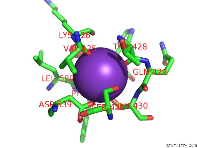
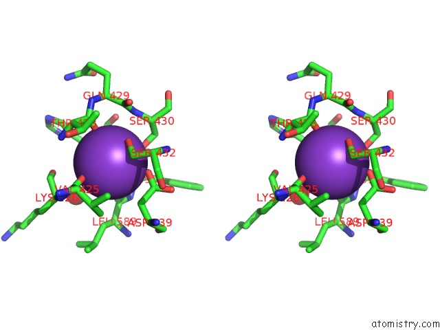
Potassium binding site 1 out of 4 in 1pkx
Go back to
Potassium binding site 1 out
of 4 in the Crystal Structure of Human Atic in Complex with Xmp
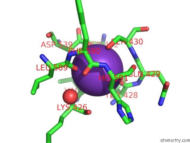
Mono view
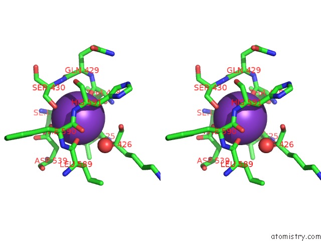
Stereo pair view
Mono view
Stereo pair view
A full contact list of Potassium with other atoms in the K binding
site number 1 of Crystal Structure of Human Atic in Complex with Xmp within 5.0Å range:
|
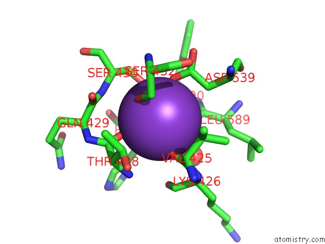
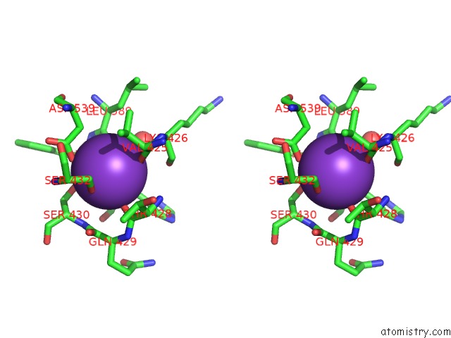
Potassium binding site 2 out of 4 in 1pkx
Go back to
Potassium binding site 2 out
of 4 in the Crystal Structure of Human Atic in Complex with Xmp
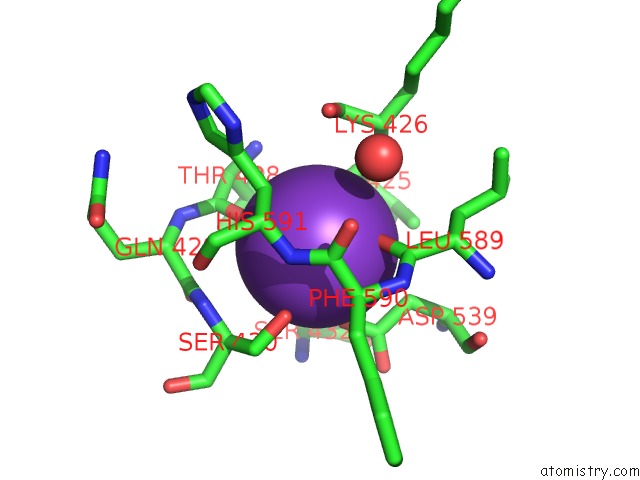
Mono view
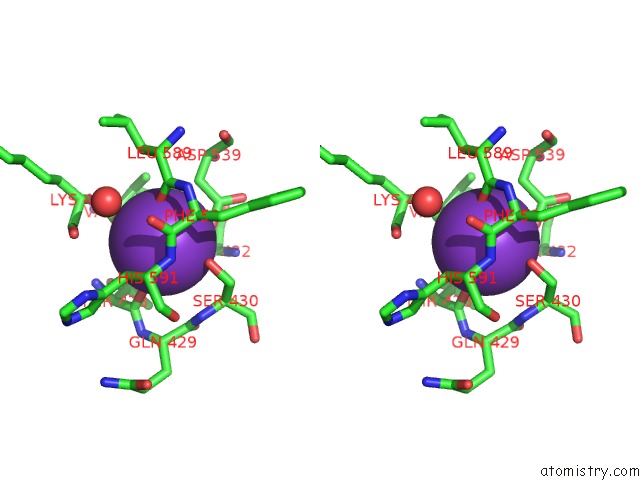
Stereo pair view
Mono view
Stereo pair view
A full contact list of Potassium with other atoms in the K binding
site number 2 of Crystal Structure of Human Atic in Complex with Xmp within 5.0Å range:
|
Potassium binding site 3 out of 4 in 1pkx
Go back to
Potassium binding site 3 out
of 4 in the Crystal Structure of Human Atic in Complex with Xmp
Mono view
Stereo pair view
Mono view
Stereo pair view
A full contact list of Potassium with other atoms in the K binding
site number 3 of Crystal Structure of Human Atic in Complex with Xmp within 5.0Å range:
|
Potassium binding site 4 out of 4 in 1pkx
Go back to
Potassium binding site 4 out
of 4 in the Crystal Structure of Human Atic in Complex with Xmp
Mono view
Stereo pair view
Mono view
Stereo pair view
A full contact list of Potassium with other atoms in the K binding
site number 4 of Crystal Structure of Human Atic in Complex with Xmp within 5.0Å range:
|
Reference:
D.W.Wolan,
C.G.Cheong,
S.E.Greasley,
I.A.Wilson.
Structural Insights Into the Human and Avian Imp Cyclohydrolase Mechanism Via Crystal Structures with the Bound Xmp Inhibitor. Biochemistry V. 43 1171 2004.
ISSN: ISSN 0006-2960
PubMed: 14756553
DOI: 10.1021/BI030162I
Page generated: Mon Aug 12 05:12:11 2024
ISSN: ISSN 0006-2960
PubMed: 14756553
DOI: 10.1021/BI030162I
Last articles
Zn in 9J0NZn in 9J0O
Zn in 9J0P
Zn in 9FJX
Zn in 9EKB
Zn in 9C0F
Zn in 9CAH
Zn in 9CH0
Zn in 9CH3
Zn in 9CH1