Potassium »
PDB 1o76-1pqo »
1p7b »
Potassium in PDB 1p7b: Crystal Structure of An Inward Rectifier Potassium Channel
Protein crystallography data
The structure of Crystal Structure of An Inward Rectifier Potassium Channel, PDB code: 1p7b
was solved by
A.Kuo,
J.M.Gulbis,
J.F.Antcliff,
T.Rahman,
E.D.Lowe,
J.Zimmer,
J.Cuthbertson,
F.M.Ashcroft,
T.Ezaki,
D.A.Doyle,
with X-Ray Crystallography technique. A brief refinement statistics is given in the table below:
| Resolution Low / High (Å) | 7.50 / 3.65 |
| Space group | I 2 2 2 |
| Cell size a, b, c (Å), α, β, γ (°) | 92.840, 105.620, 258.630, 90.00, 90.00, 90.00 |
| R / Rfree (%) | 29.5 / 32.9 |
Potassium Binding Sites:
The binding sites of Potassium atom in the Crystal Structure of An Inward Rectifier Potassium Channel
(pdb code 1p7b). This binding sites where shown within
5.0 Angstroms radius around Potassium atom.
In total 4 binding sites of Potassium where determined in the Crystal Structure of An Inward Rectifier Potassium Channel, PDB code: 1p7b:
Jump to Potassium binding site number: 1; 2; 3; 4;
In total 4 binding sites of Potassium where determined in the Crystal Structure of An Inward Rectifier Potassium Channel, PDB code: 1p7b:
Jump to Potassium binding site number: 1; 2; 3; 4;
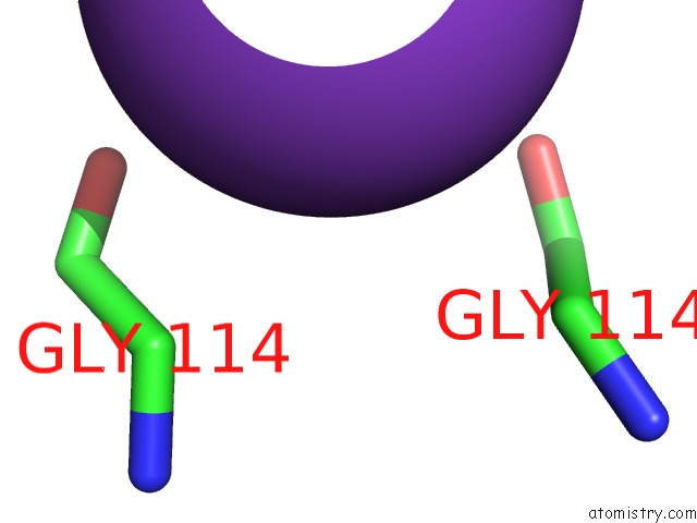
Potassium binding site 1 out of 4 in 1p7b
Go back to
Potassium binding site 1 out
of 4 in the Crystal Structure of An Inward Rectifier Potassium Channel
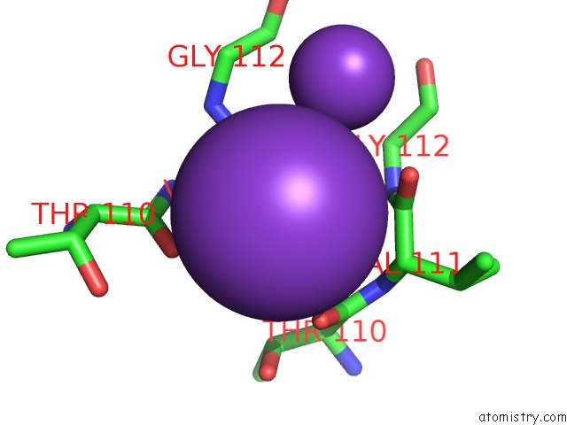
Mono view
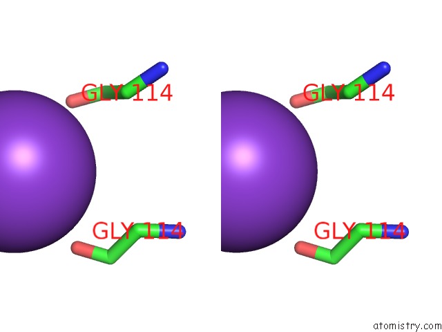
Stereo pair view
Mono view
Stereo pair view
A full contact list of Potassium with other atoms in the K binding
site number 1 of Crystal Structure of An Inward Rectifier Potassium Channel within 5.0Å range:
|
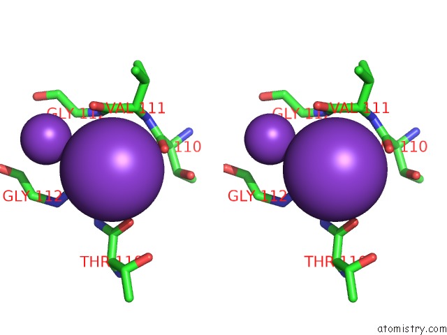
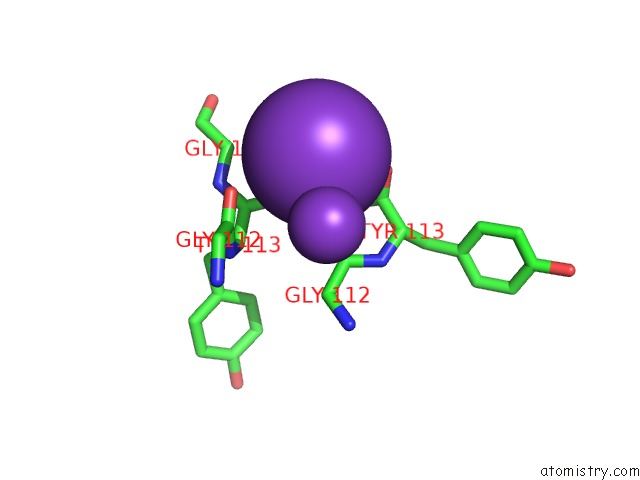
Potassium binding site 2 out of 4 in 1p7b
Go back to
Potassium binding site 2 out
of 4 in the Crystal Structure of An Inward Rectifier Potassium Channel
Mono view
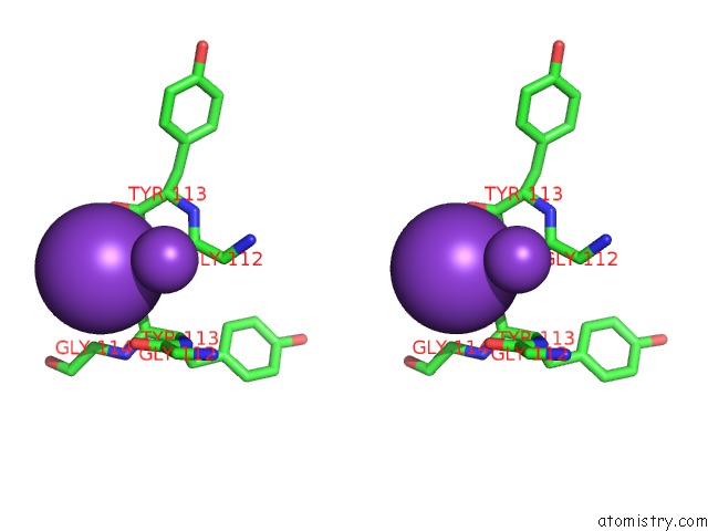
Stereo pair view
Mono view
Stereo pair view
A full contact list of Potassium with other atoms in the K binding
site number 2 of Crystal Structure of An Inward Rectifier Potassium Channel within 5.0Å range:
|
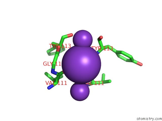
Potassium binding site 3 out of 4 in 1p7b
Go back to
Potassium binding site 3 out
of 4 in the Crystal Structure of An Inward Rectifier Potassium Channel
Mono view
Stereo pair view
Mono view
Stereo pair view
A full contact list of Potassium with other atoms in the K binding
site number 3 of Crystal Structure of An Inward Rectifier Potassium Channel within 5.0Å range:
|
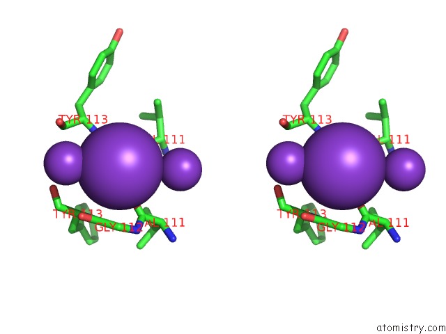
Potassium binding site 4 out of 4 in 1p7b
Go back to
Potassium binding site 4 out
of 4 in the Crystal Structure of An Inward Rectifier Potassium Channel
Mono view
Stereo pair view
Mono view
Stereo pair view
A full contact list of Potassium with other atoms in the K binding
site number 4 of Crystal Structure of An Inward Rectifier Potassium Channel within 5.0Å range:
|
Reference:
A.Kuo,
J.M.Gulbis,
J.F.Antcliff,
T.Rahman,
E.D.Lowe,
J.Zimmer,
J.Cuthbertson,
F.M.Ashcroft,
T.Ezaki,
D.A.Doyle.
Crystal Structure of the Potassium Channel KIRBAC1.1 in the Closed State. Science V. 300 1922 2003.
ISSN: ISSN 0036-8075
PubMed: 12738871
DOI: 10.1126/SCIENCE.1085028
Page generated: Mon Aug 12 05:10:10 2024
ISSN: ISSN 0036-8075
PubMed: 12738871
DOI: 10.1126/SCIENCE.1085028
Last articles
Zn in 9J0NZn in 9J0O
Zn in 9J0P
Zn in 9FJX
Zn in 9EKB
Zn in 9C0F
Zn in 9CAH
Zn in 9CH0
Zn in 9CH3
Zn in 9CH1