Potassium »
PDB 1o76-1pqo »
1o92 »
Potassium in PDB 1o92: Methionine Adenosyltransferase Complexed with Adp and A L-Methionine Analogue
Enzymatic activity of Methionine Adenosyltransferase Complexed with Adp and A L-Methionine Analogue
All present enzymatic activity of Methionine Adenosyltransferase Complexed with Adp and A L-Methionine Analogue:
2.5.1.6;
2.5.1.6;
Protein crystallography data
The structure of Methionine Adenosyltransferase Complexed with Adp and A L-Methionine Analogue, PDB code: 1o92
was solved by
B.Gonzalez,
M.A.Pajares,
J.A.Hermoso,
J.Sanz-Aparicio,
with X-Ray Crystallography technique. A brief refinement statistics is given in the table below:
| Resolution Low / High (Å) | 8.00 / 3.19 |
| Space group | P 41 2 2 |
| Cell size a, b, c (Å), α, β, γ (°) | 115.140, 115.140, 160.450, 90.00, 90.00, 90.00 |
| R / Rfree (%) | 24.6 / 26.4 |
Other elements in 1o92:
The structure of Methionine Adenosyltransferase Complexed with Adp and A L-Methionine Analogue also contains other interesting chemical elements:
| Magnesium | (Mg) | 3 atoms |
Potassium Binding Sites:
The binding sites of Potassium atom in the Methionine Adenosyltransferase Complexed with Adp and A L-Methionine Analogue
(pdb code 1o92). This binding sites where shown within
5.0 Angstroms radius around Potassium atom.
In total 2 binding sites of Potassium where determined in the Methionine Adenosyltransferase Complexed with Adp and A L-Methionine Analogue, PDB code: 1o92:
Jump to Potassium binding site number: 1; 2;
In total 2 binding sites of Potassium where determined in the Methionine Adenosyltransferase Complexed with Adp and A L-Methionine Analogue, PDB code: 1o92:
Jump to Potassium binding site number: 1; 2;
Potassium binding site 1 out of 2 in 1o92
Go back to
Potassium binding site 1 out
of 2 in the Methionine Adenosyltransferase Complexed with Adp and A L-Methionine Analogue
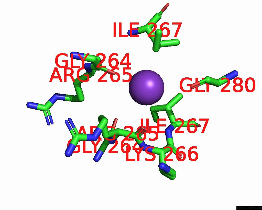
Mono view
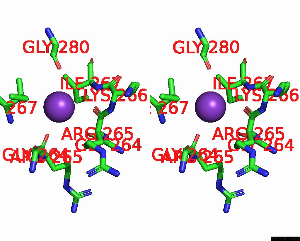
Stereo pair view
Mono view
Stereo pair view
A full contact list of Potassium with other atoms in the K binding
site number 1 of Methionine Adenosyltransferase Complexed with Adp and A L-Methionine Analogue within 5.0Å range:
|
Potassium binding site 2 out of 2 in 1o92
Go back to
Potassium binding site 2 out
of 2 in the Methionine Adenosyltransferase Complexed with Adp and A L-Methionine Analogue
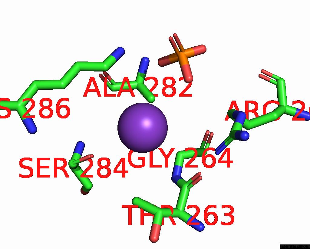
Mono view
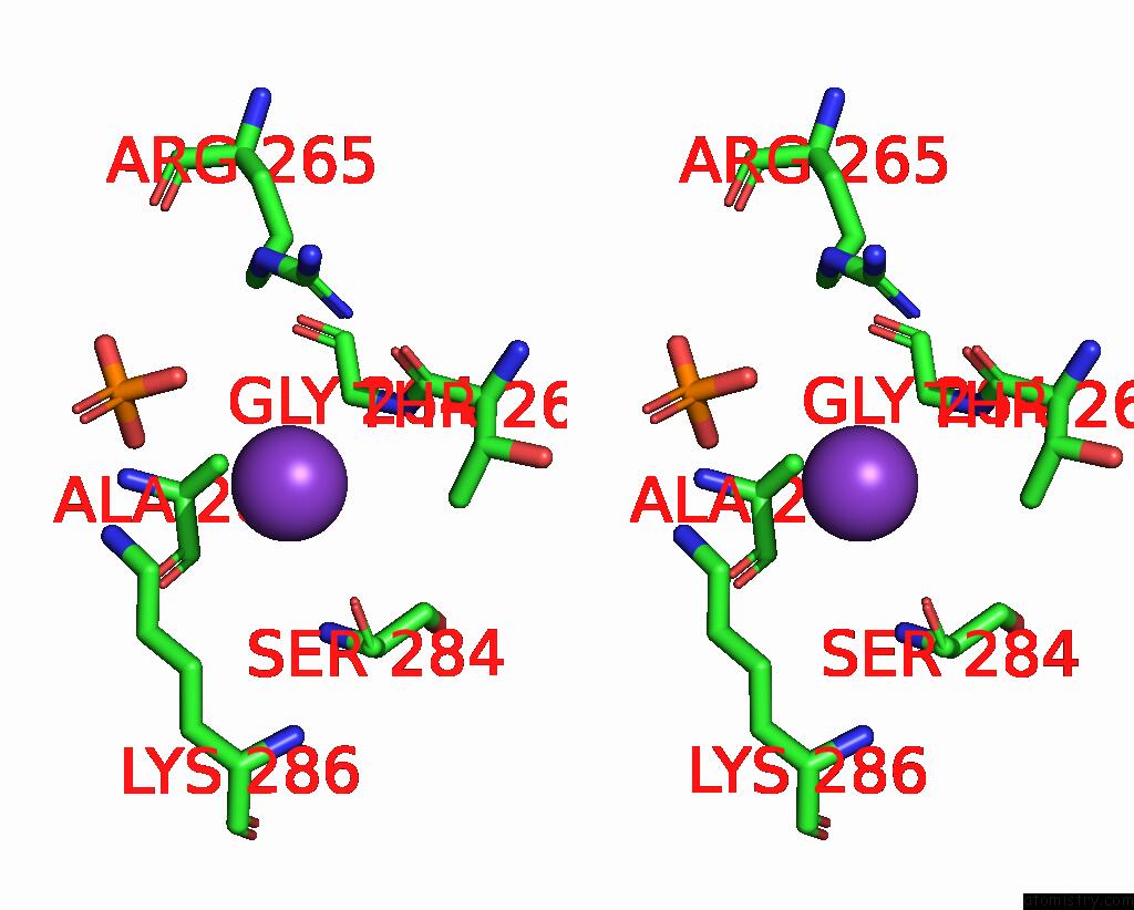
Stereo pair view
Mono view
Stereo pair view
A full contact list of Potassium with other atoms in the K binding
site number 2 of Methionine Adenosyltransferase Complexed with Adp and A L-Methionine Analogue within 5.0Å range:
|
Reference:
B.Gonzalez,
M.A.Pajares,
J.A.Hermoso,
D.Guillerm,
G.Guillerm,
J.Sanz-Aparicio.
Crystal Structures of Methionine Adenosyltransferase Complexed with Substrates and Products Reveal the Methionine-Atp Recognition and Give Insights Into the Catalytic Mechanism J.Mol.Biol. V. 331 407 2003.
ISSN: ISSN 0022-2836
PubMed: 12888348
DOI: 10.1016/S0022-2836(03)00728-9
Page generated: Mon Aug 12 05:06:48 2024
ISSN: ISSN 0022-2836
PubMed: 12888348
DOI: 10.1016/S0022-2836(03)00728-9
Last articles
Zn in 9J0NZn in 9J0O
Zn in 9J0P
Zn in 9FJX
Zn in 9EKB
Zn in 9C0F
Zn in 9CAH
Zn in 9CH0
Zn in 9CH3
Zn in 9CH1