Potassium »
PDB 1o76-1pqo »
1o76 »
Potassium in PDB 1o76: Cyanide Complex of P450CAM From Pseudomonas Putida
Enzymatic activity of Cyanide Complex of P450CAM From Pseudomonas Putida
All present enzymatic activity of Cyanide Complex of P450CAM From Pseudomonas Putida:
1.14.15.1;
1.14.15.1;
Protein crystallography data
The structure of Cyanide Complex of P450CAM From Pseudomonas Putida, PDB code: 1o76
was solved by
R.Fedorov,
D.Ghosh,
I.Schlichting,
with X-Ray Crystallography technique. A brief refinement statistics is given in the table below:
| Resolution Low / High (Å) | 19.00 / 1.80 |
| Space group | P 1 21 1 |
| Cell size a, b, c (Å), α, β, γ (°) | 67.000, 61.800, 94.600, 90.00, 90.40, 90.00 |
| R / Rfree (%) | 19.6 / 23.6 |
Other elements in 1o76:
The structure of Cyanide Complex of P450CAM From Pseudomonas Putida also contains other interesting chemical elements:
| Iron | (Fe) | 2 atoms |
Potassium Binding Sites:
The binding sites of Potassium atom in the Cyanide Complex of P450CAM From Pseudomonas Putida
(pdb code 1o76). This binding sites where shown within
5.0 Angstroms radius around Potassium atom.
In total 3 binding sites of Potassium where determined in the Cyanide Complex of P450CAM From Pseudomonas Putida, PDB code: 1o76:
Jump to Potassium binding site number: 1; 2; 3;
In total 3 binding sites of Potassium where determined in the Cyanide Complex of P450CAM From Pseudomonas Putida, PDB code: 1o76:
Jump to Potassium binding site number: 1; 2; 3;
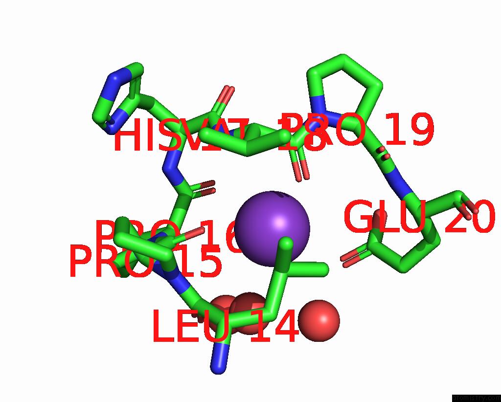
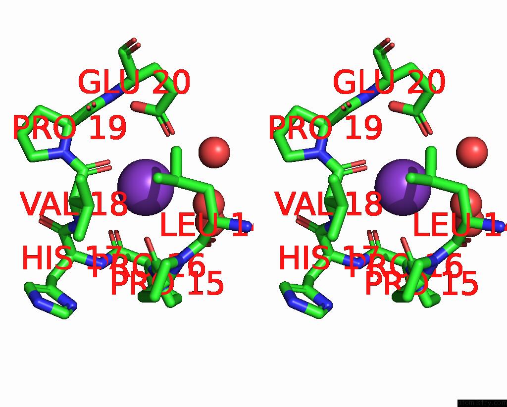
Potassium binding site 1 out of 3 in 1o76
Go back to
Potassium binding site 1 out
of 3 in the Cyanide Complex of P450CAM From Pseudomonas Putida
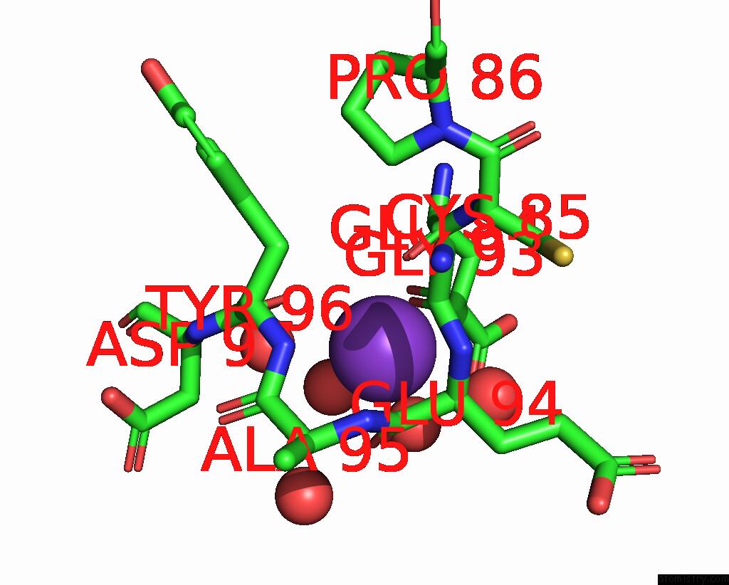
Mono view
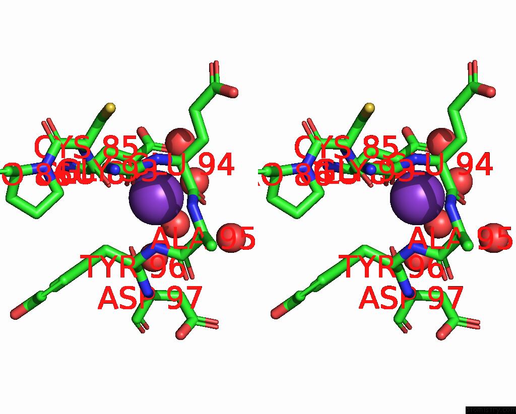
Stereo pair view
Mono view
Stereo pair view
A full contact list of Potassium with other atoms in the K binding
site number 1 of Cyanide Complex of P450CAM From Pseudomonas Putida within 5.0Å range:
|
Potassium binding site 2 out of 3 in 1o76
Go back to
Potassium binding site 2 out
of 3 in the Cyanide Complex of P450CAM From Pseudomonas Putida
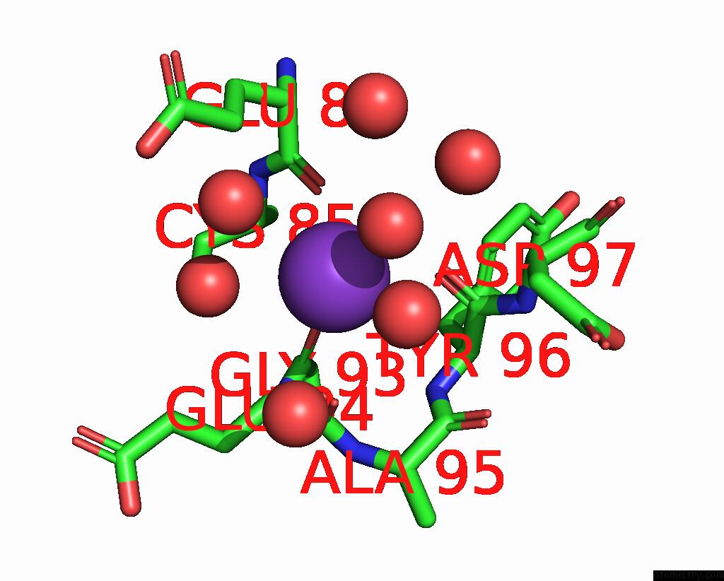
Mono view
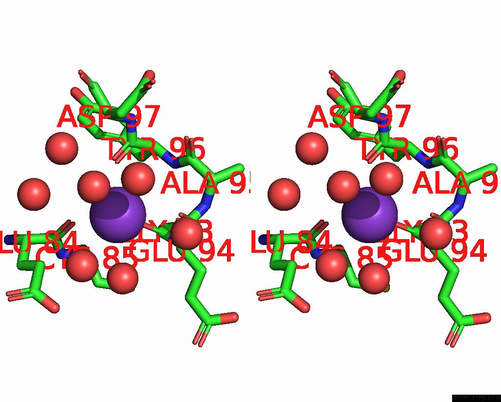
Stereo pair view
Mono view
Stereo pair view
A full contact list of Potassium with other atoms in the K binding
site number 2 of Cyanide Complex of P450CAM From Pseudomonas Putida within 5.0Å range:
|
Potassium binding site 3 out of 3 in 1o76
Go back to
Potassium binding site 3 out
of 3 in the Cyanide Complex of P450CAM From Pseudomonas Putida
Mono view
Stereo pair view
Mono view
Stereo pair view
A full contact list of Potassium with other atoms in the K binding
site number 3 of Cyanide Complex of P450CAM From Pseudomonas Putida within 5.0Å range:
|
Reference:
R.Fedorov,
D.Ghosh,
I.Schlichting.
Crystal Structures of Cyanide Complexes of P450CAM and the Oxygenase Domain of Inducible Nitric Oxide Synthase-Structural Models of the Short-Lived Oxygen Complexes Arch.Biochem.Biophys. V. 409 25 2003.
ISSN: ISSN 0003-9861
PubMed: 12464241
DOI: 10.1016/S0003-9861(02)00555-6
Page generated: Mon Aug 12 05:06:48 2024
ISSN: ISSN 0003-9861
PubMed: 12464241
DOI: 10.1016/S0003-9861(02)00555-6
Last articles
Zn in 9J0NZn in 9J0O
Zn in 9J0P
Zn in 9FJX
Zn in 9EKB
Zn in 9C0F
Zn in 9CAH
Zn in 9CH0
Zn in 9CH3
Zn in 9CH1