Potassium »
PDB 1m5h-1o07 »
1o07 »
Potassium in PDB 1o07: Crystal Structure of the Complex Between Q120L/Y150E Mutant of Ampc and A Beta-Lactam Inhibitor (Mxg)
Enzymatic activity of Crystal Structure of the Complex Between Q120L/Y150E Mutant of Ampc and A Beta-Lactam Inhibitor (Mxg)
All present enzymatic activity of Crystal Structure of the Complex Between Q120L/Y150E Mutant of Ampc and A Beta-Lactam Inhibitor (Mxg):
3.5.2.6;
3.5.2.6;
Protein crystallography data
The structure of Crystal Structure of the Complex Between Q120L/Y150E Mutant of Ampc and A Beta-Lactam Inhibitor (Mxg), PDB code: 1o07
was solved by
S.O.Meroueh,
G.Minasov,
W.Lee,
B.K.Shoichet,
S.Mobashery,
with X-Ray Crystallography technique. A brief refinement statistics is given in the table below:
| Resolution Low / High (Å) | 24.77 / 1.71 |
| Space group | C 1 2 1 |
| Cell size a, b, c (Å), α, β, γ (°) | 118.808, 76.298, 98.127, 90.00, 116.21, 90.00 |
| R / Rfree (%) | 15.4 / 19.1 |
Potassium Binding Sites:
The binding sites of Potassium atom in the Crystal Structure of the Complex Between Q120L/Y150E Mutant of Ampc and A Beta-Lactam Inhibitor (Mxg)
(pdb code 1o07). This binding sites where shown within
5.0 Angstroms radius around Potassium atom.
In total 2 binding sites of Potassium where determined in the Crystal Structure of the Complex Between Q120L/Y150E Mutant of Ampc and A Beta-Lactam Inhibitor (Mxg), PDB code: 1o07:
Jump to Potassium binding site number: 1; 2;
In total 2 binding sites of Potassium where determined in the Crystal Structure of the Complex Between Q120L/Y150E Mutant of Ampc and A Beta-Lactam Inhibitor (Mxg), PDB code: 1o07:
Jump to Potassium binding site number: 1; 2;
Potassium binding site 1 out of 2 in 1o07
Go back to
Potassium binding site 1 out
of 2 in the Crystal Structure of the Complex Between Q120L/Y150E Mutant of Ampc and A Beta-Lactam Inhibitor (Mxg)
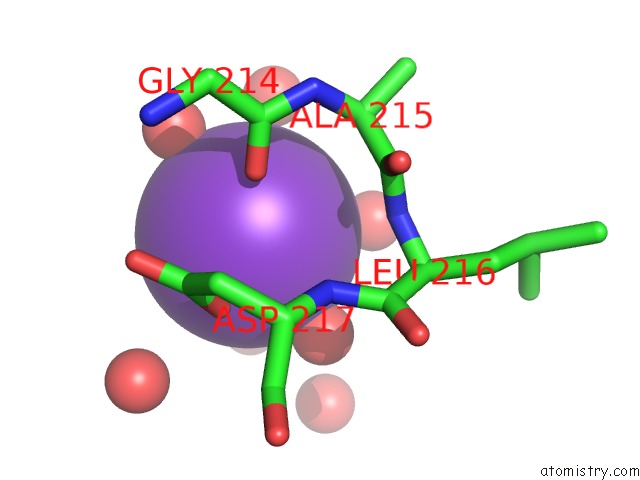
Mono view
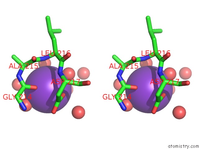
Stereo pair view
Mono view
Stereo pair view
A full contact list of Potassium with other atoms in the K binding
site number 1 of Crystal Structure of the Complex Between Q120L/Y150E Mutant of Ampc and A Beta-Lactam Inhibitor (Mxg) within 5.0Å range:
|
Potassium binding site 2 out of 2 in 1o07
Go back to
Potassium binding site 2 out
of 2 in the Crystal Structure of the Complex Between Q120L/Y150E Mutant of Ampc and A Beta-Lactam Inhibitor (Mxg)
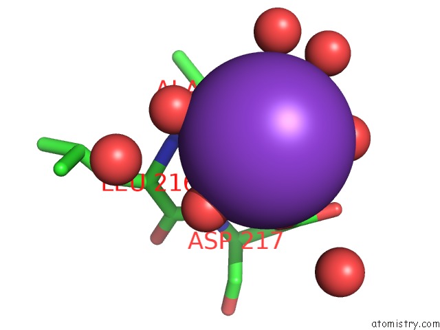
Mono view
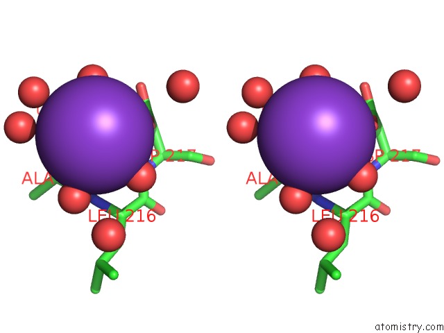
Stereo pair view
Mono view
Stereo pair view
A full contact list of Potassium with other atoms in the K binding
site number 2 of Crystal Structure of the Complex Between Q120L/Y150E Mutant of Ampc and A Beta-Lactam Inhibitor (Mxg) within 5.0Å range:
|
Reference:
S.O.Meroueh,
G.Minasov,
W.Lee,
B.K.Shoichet,
S.Mobashery.
Structural Aspects For Evolution of Beta-Lactamases From Penicillin-Binding Proteins J.Am.Chem.Soc. V. 125 9612 2003.
ISSN: ISSN 0002-7863
PubMed: 12904027
DOI: 10.1021/JA034861U
Page generated: Mon Aug 12 05:04:54 2024
ISSN: ISSN 0002-7863
PubMed: 12904027
DOI: 10.1021/JA034861U
Last articles
Zn in 9J0NZn in 9J0O
Zn in 9J0P
Zn in 9FJX
Zn in 9EKB
Zn in 9C0F
Zn in 9CAH
Zn in 9CH0
Zn in 9CH3
Zn in 9CH1