Potassium »
PDB 1m5h-1o07 »
1mew »
Potassium in PDB 1mew: Inosine Monophosphate Dehydrogenase (Impdh) From Tritrichomonas Foetus with Xmp and Nad Bound
Enzymatic activity of Inosine Monophosphate Dehydrogenase (Impdh) From Tritrichomonas Foetus with Xmp and Nad Bound
All present enzymatic activity of Inosine Monophosphate Dehydrogenase (Impdh) From Tritrichomonas Foetus with Xmp and Nad Bound:
1.1.1.205;
1.1.1.205;
Protein crystallography data
The structure of Inosine Monophosphate Dehydrogenase (Impdh) From Tritrichomonas Foetus with Xmp and Nad Bound, PDB code: 1mew
was solved by
G.L.Prosise,
H.Luecke,
with X-Ray Crystallography technique. A brief refinement statistics is given in the table below:
| Resolution Low / High (Å) | 29.61 / 2.15 |
| Space group | P 4 3 2 |
| Cell size a, b, c (Å), α, β, γ (°) | 153.827, 153.827, 153.827, 90.00, 90.00, 90.00 |
| R / Rfree (%) | 22.4 / 24.6 |
Potassium Binding Sites:
The binding sites of Potassium atom in the Inosine Monophosphate Dehydrogenase (Impdh) From Tritrichomonas Foetus with Xmp and Nad Bound
(pdb code 1mew). This binding sites where shown within
5.0 Angstroms radius around Potassium atom.
In total only one binding site of Potassium was determined in the Inosine Monophosphate Dehydrogenase (Impdh) From Tritrichomonas Foetus with Xmp and Nad Bound, PDB code: 1mew:
In total only one binding site of Potassium was determined in the Inosine Monophosphate Dehydrogenase (Impdh) From Tritrichomonas Foetus with Xmp and Nad Bound, PDB code: 1mew:
Potassium binding site 1 out of 1 in 1mew
Go back to
Potassium binding site 1 out
of 1 in the Inosine Monophosphate Dehydrogenase (Impdh) From Tritrichomonas Foetus with Xmp and Nad Bound
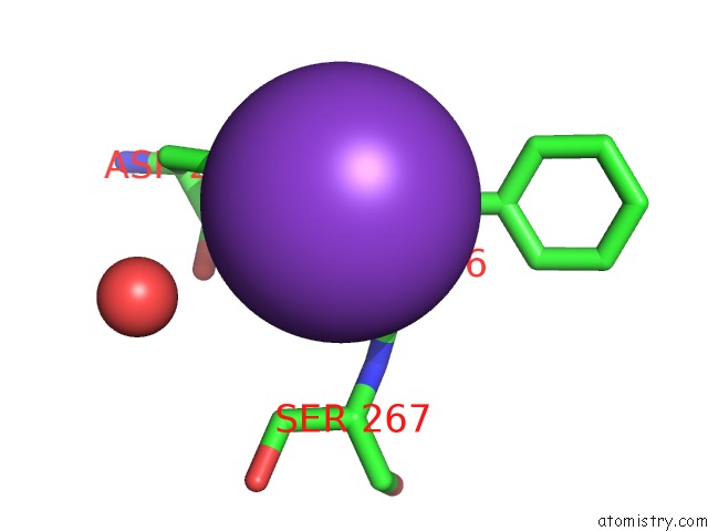
Mono view
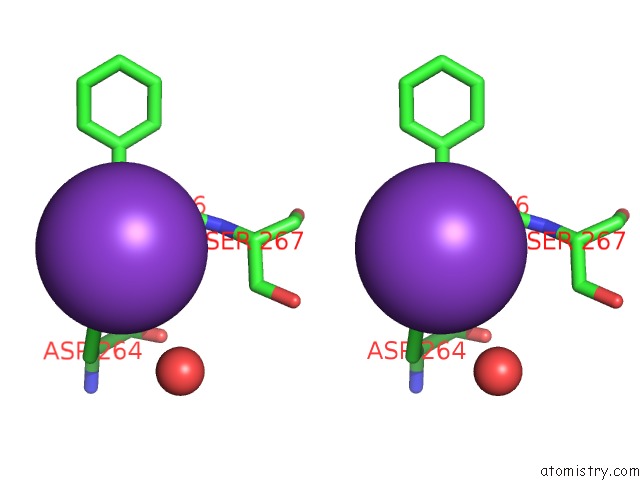
Stereo pair view
Mono view
Stereo pair view
A full contact list of Potassium with other atoms in the K binding
site number 1 of Inosine Monophosphate Dehydrogenase (Impdh) From Tritrichomonas Foetus with Xmp and Nad Bound within 5.0Å range:
|
Reference:
G.L.Prosise,
H.Luecke.
Crystal Structures of Tritrichomonas Foetus Inosine Monophosphate Dehydrogenase in Complex with Substrate, Cofactor and Analogs: A Structural Basis For the Random-in Ordered-Out Kinetic Mechanism J.Mol.Biol. V. 326 517 2003.
ISSN: ISSN 0022-2836
PubMed: 12559919
DOI: 10.1016/S0022-2836(02)01383-9
Page generated: Mon Aug 12 04:57:56 2024
ISSN: ISSN 0022-2836
PubMed: 12559919
DOI: 10.1016/S0022-2836(02)01383-9
Last articles
Zn in 9J0NZn in 9J0O
Zn in 9J0P
Zn in 9FJX
Zn in 9EKB
Zn in 9C0F
Zn in 9CAH
Zn in 9CH0
Zn in 9CH3
Zn in 9CH1