Potassium »
PDB 1m5h-1o07 »
1m9n »
Potassium in PDB 1m9n: Crystal Structure of the Homodimeric Bifunctional Transformylase and Cyclohydrolase Enzyme Avian Atic in Complex with Aicar and Xmp at 1.93 Angstroms.
Enzymatic activity of Crystal Structure of the Homodimeric Bifunctional Transformylase and Cyclohydrolase Enzyme Avian Atic in Complex with Aicar and Xmp at 1.93 Angstroms.
All present enzymatic activity of Crystal Structure of the Homodimeric Bifunctional Transformylase and Cyclohydrolase Enzyme Avian Atic in Complex with Aicar and Xmp at 1.93 Angstroms.:
2.1.2.3; 3.5.4.10;
2.1.2.3; 3.5.4.10;
Protein crystallography data
The structure of Crystal Structure of the Homodimeric Bifunctional Transformylase and Cyclohydrolase Enzyme Avian Atic in Complex with Aicar and Xmp at 1.93 Angstroms., PDB code: 1m9n
was solved by
D.W.Wolan,
S.E.Greasly,
G.P.Beardsley,
I.A.Wilson,
with X-Ray Crystallography technique. A brief refinement statistics is given in the table below:
| Resolution Low / High (Å) | 29.24 / 1.93 |
| Space group | P 1 21 1 |
| Cell size a, b, c (Å), α, β, γ (°) | 56.480, 107.880, 103.860, 90.00, 91.20, 90.00 |
| R / Rfree (%) | 20.6 / 24.4 |
Potassium Binding Sites:
The binding sites of Potassium atom in the Crystal Structure of the Homodimeric Bifunctional Transformylase and Cyclohydrolase Enzyme Avian Atic in Complex with Aicar and Xmp at 1.93 Angstroms.
(pdb code 1m9n). This binding sites where shown within
5.0 Angstroms radius around Potassium atom.
In total 2 binding sites of Potassium where determined in the Crystal Structure of the Homodimeric Bifunctional Transformylase and Cyclohydrolase Enzyme Avian Atic in Complex with Aicar and Xmp at 1.93 Angstroms., PDB code: 1m9n:
Jump to Potassium binding site number: 1; 2;
In total 2 binding sites of Potassium where determined in the Crystal Structure of the Homodimeric Bifunctional Transformylase and Cyclohydrolase Enzyme Avian Atic in Complex with Aicar and Xmp at 1.93 Angstroms., PDB code: 1m9n:
Jump to Potassium binding site number: 1; 2;
Potassium binding site 1 out of 2 in 1m9n
Go back to
Potassium binding site 1 out
of 2 in the Crystal Structure of the Homodimeric Bifunctional Transformylase and Cyclohydrolase Enzyme Avian Atic in Complex with Aicar and Xmp at 1.93 Angstroms.
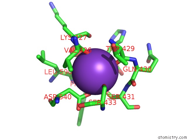
Mono view
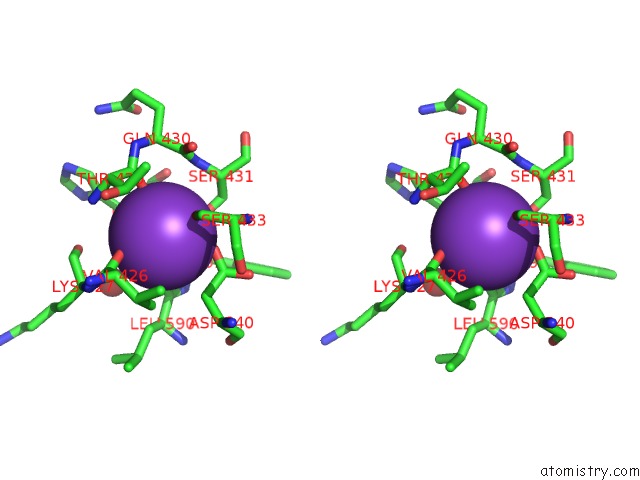
Stereo pair view
Mono view
Stereo pair view
A full contact list of Potassium with other atoms in the K binding
site number 1 of Crystal Structure of the Homodimeric Bifunctional Transformylase and Cyclohydrolase Enzyme Avian Atic in Complex with Aicar and Xmp at 1.93 Angstroms. within 5.0Å range:
|
Potassium binding site 2 out of 2 in 1m9n
Go back to
Potassium binding site 2 out
of 2 in the Crystal Structure of the Homodimeric Bifunctional Transformylase and Cyclohydrolase Enzyme Avian Atic in Complex with Aicar and Xmp at 1.93 Angstroms.
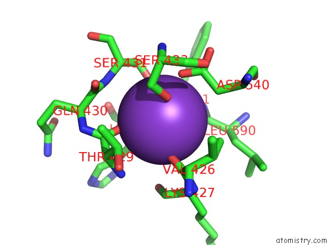
Mono view
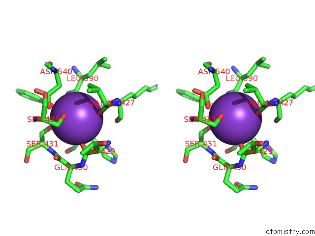
Stereo pair view
Mono view
Stereo pair view
A full contact list of Potassium with other atoms in the K binding
site number 2 of Crystal Structure of the Homodimeric Bifunctional Transformylase and Cyclohydrolase Enzyme Avian Atic in Complex with Aicar and Xmp at 1.93 Angstroms. within 5.0Å range:
|
Reference:
D.W.Wolan,
S.E.Greasly,
G.P.Beardsley,
I.A.Wilson.
Structural Insights Into the Avian Aicar Transformylase Mechanism. Biochemistry V. 41 15505 2002.
ISSN: ISSN 0006-2960
PubMed: 12501179
DOI: 10.1021/BI020505X
Page generated: Mon Aug 12 04:56:07 2024
ISSN: ISSN 0006-2960
PubMed: 12501179
DOI: 10.1021/BI020505X
Last articles
Zn in 9J0NZn in 9J0O
Zn in 9J0P
Zn in 9FJX
Zn in 9EKB
Zn in 9C0F
Zn in 9CAH
Zn in 9CH0
Zn in 9CH3
Zn in 9CH1