Potassium »
PDB 1k4d-1m40 »
1lvg »
Potassium in PDB 1lvg: Crystal Structure of Mouse Guanylate Kinase in Complex with Gmp and Adp
Enzymatic activity of Crystal Structure of Mouse Guanylate Kinase in Complex with Gmp and Adp
All present enzymatic activity of Crystal Structure of Mouse Guanylate Kinase in Complex with Gmp and Adp:
2.7.4.8;
2.7.4.8;
Protein crystallography data
The structure of Crystal Structure of Mouse Guanylate Kinase in Complex with Gmp and Adp, PDB code: 1lvg
was solved by
N.Sekulic,
L.Shuvalova,
O.Spangenberg,
M.Konrad,
A.Lavie,
with X-Ray Crystallography technique. A brief refinement statistics is given in the table below:
| Resolution Low / High (Å) | 20.00 / 2.10 |
| Space group | P 41 21 2 |
| Cell size a, b, c (Å), α, β, γ (°) | 67.241, 67.241, 108.696, 90.00, 90.00, 90.00 |
| R / Rfree (%) | 18.8 / 23.1 |
Potassium Binding Sites:
The binding sites of Potassium atom in the Crystal Structure of Mouse Guanylate Kinase in Complex with Gmp and Adp
(pdb code 1lvg). This binding sites where shown within
5.0 Angstroms radius around Potassium atom.
In total only one binding site of Potassium was determined in the Crystal Structure of Mouse Guanylate Kinase in Complex with Gmp and Adp, PDB code: 1lvg:
In total only one binding site of Potassium was determined in the Crystal Structure of Mouse Guanylate Kinase in Complex with Gmp and Adp, PDB code: 1lvg:
Potassium binding site 1 out of 1 in 1lvg
Go back to
Potassium binding site 1 out
of 1 in the Crystal Structure of Mouse Guanylate Kinase in Complex with Gmp and Adp
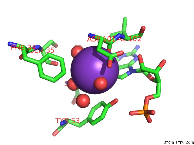
Mono view
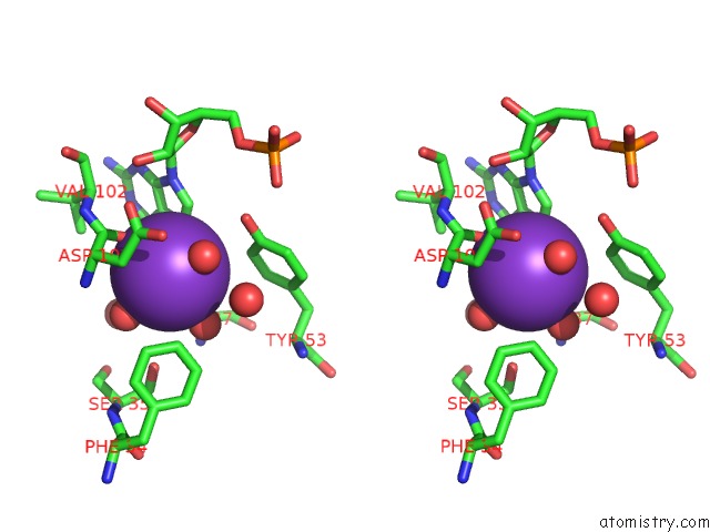
Stereo pair view
Mono view
Stereo pair view
A full contact list of Potassium with other atoms in the K binding
site number 1 of Crystal Structure of Mouse Guanylate Kinase in Complex with Gmp and Adp within 5.0Å range:
|
Reference:
N.Sekulic,
L.Shuvalova,
O.Spangenberg,
M.Konrad,
A.Lavie.
Structural Characterization of the Closed Conformation of Mouse Guanylate Kinase. J.Biol.Chem. V. 277 30236 2002.
ISSN: ISSN 0021-9258
PubMed: 12036965
DOI: 10.1074/JBC.M204668200
Page generated: Mon Aug 12 04:52:37 2024
ISSN: ISSN 0021-9258
PubMed: 12036965
DOI: 10.1074/JBC.M204668200
Last articles
Zn in 9J0NZn in 9J0O
Zn in 9J0P
Zn in 9FJX
Zn in 9EKB
Zn in 9C0F
Zn in 9CAH
Zn in 9CH0
Zn in 9CH3
Zn in 9CH1