Potassium »
PDB 1gup-1k4c »
1jvj »
Potassium in PDB 1jvj: Crystal Structure of N132A Mutant of Tem-1 Beta-Lactamase in Complex with A N-Formimidoyl-Thienamycine
Enzymatic activity of Crystal Structure of N132A Mutant of Tem-1 Beta-Lactamase in Complex with A N-Formimidoyl-Thienamycine
All present enzymatic activity of Crystal Structure of N132A Mutant of Tem-1 Beta-Lactamase in Complex with A N-Formimidoyl-Thienamycine:
3.5.2.6;
3.5.2.6;
Protein crystallography data
The structure of Crystal Structure of N132A Mutant of Tem-1 Beta-Lactamase in Complex with A N-Formimidoyl-Thienamycine, PDB code: 1jvj
was solved by
X.Wang,
G.Minasov,
B.K.Shoichet,
with X-Ray Crystallography technique. A brief refinement statistics is given in the table below:
| Resolution Low / High (Å) | 15.00 / 1.73 |
| Space group | P 21 21 21 |
| Cell size a, b, c (Å), α, β, γ (°) | 41.303, 61.685, 89.139, 90.00, 90.00, 90.00 |
| R / Rfree (%) | 16.2 / 19.3 |
Potassium Binding Sites:
The binding sites of Potassium atom in the Crystal Structure of N132A Mutant of Tem-1 Beta-Lactamase in Complex with A N-Formimidoyl-Thienamycine
(pdb code 1jvj). This binding sites where shown within
5.0 Angstroms radius around Potassium atom.
In total 5 binding sites of Potassium where determined in the Crystal Structure of N132A Mutant of Tem-1 Beta-Lactamase in Complex with A N-Formimidoyl-Thienamycine, PDB code: 1jvj:
Jump to Potassium binding site number: 1; 2; 3; 4; 5;
In total 5 binding sites of Potassium where determined in the Crystal Structure of N132A Mutant of Tem-1 Beta-Lactamase in Complex with A N-Formimidoyl-Thienamycine, PDB code: 1jvj:
Jump to Potassium binding site number: 1; 2; 3; 4; 5;
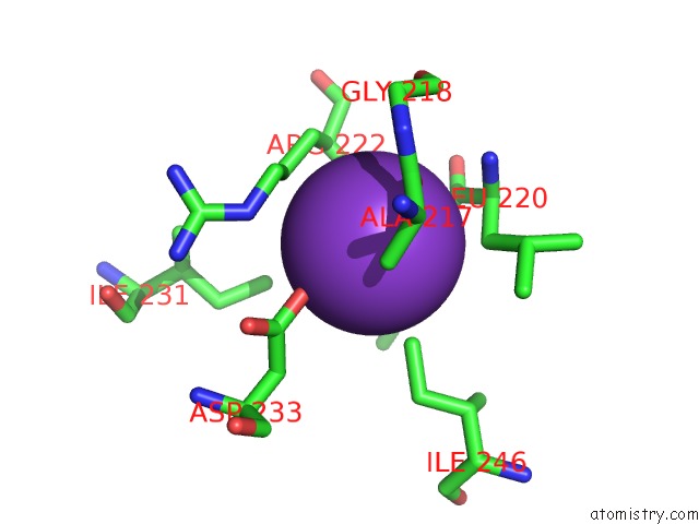
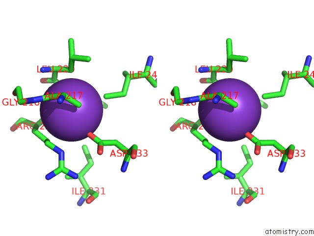
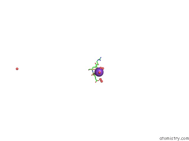
Potassium binding site 1 out of 5 in 1jvj
Go back to
Potassium binding site 1 out
of 5 in the Crystal Structure of N132A Mutant of Tem-1 Beta-Lactamase in Complex with A N-Formimidoyl-Thienamycine
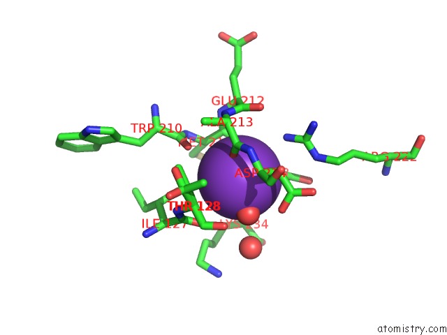
Mono view
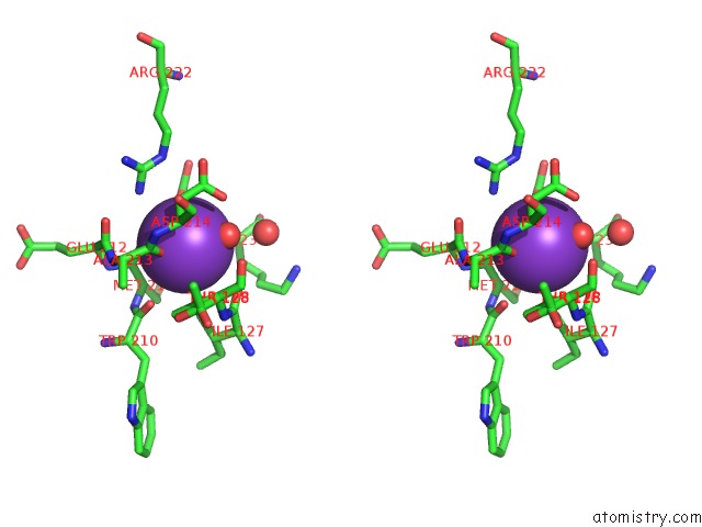
Stereo pair view
Mono view
Stereo pair view
A full contact list of Potassium with other atoms in the K binding
site number 1 of Crystal Structure of N132A Mutant of Tem-1 Beta-Lactamase in Complex with A N-Formimidoyl-Thienamycine within 5.0Å range:
|
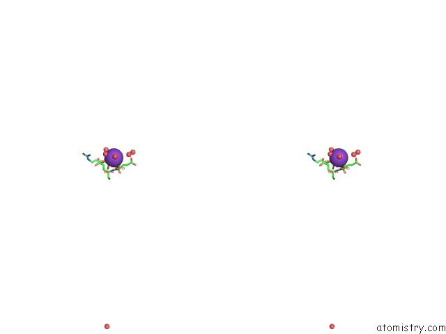
Potassium binding site 2 out of 5 in 1jvj
Go back to
Potassium binding site 2 out
of 5 in the Crystal Structure of N132A Mutant of Tem-1 Beta-Lactamase in Complex with A N-Formimidoyl-Thienamycine
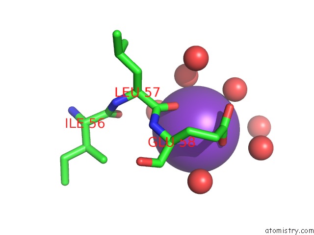
Mono view
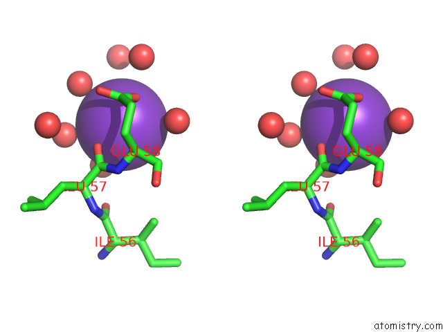
Stereo pair view
Mono view
Stereo pair view
A full contact list of Potassium with other atoms in the K binding
site number 2 of Crystal Structure of N132A Mutant of Tem-1 Beta-Lactamase in Complex with A N-Formimidoyl-Thienamycine within 5.0Å range:
|
Potassium binding site 3 out of 5 in 1jvj
Go back to
Potassium binding site 3 out
of 5 in the Crystal Structure of N132A Mutant of Tem-1 Beta-Lactamase in Complex with A N-Formimidoyl-Thienamycine
Mono view
Stereo pair view
Mono view
Stereo pair view
A full contact list of Potassium with other atoms in the K binding
site number 3 of Crystal Structure of N132A Mutant of Tem-1 Beta-Lactamase in Complex with A N-Formimidoyl-Thienamycine within 5.0Å range:
|
Potassium binding site 4 out of 5 in 1jvj
Go back to
Potassium binding site 4 out
of 5 in the Crystal Structure of N132A Mutant of Tem-1 Beta-Lactamase in Complex with A N-Formimidoyl-Thienamycine
Mono view
Stereo pair view
Mono view
Stereo pair view
A full contact list of Potassium with other atoms in the K binding
site number 4 of Crystal Structure of N132A Mutant of Tem-1 Beta-Lactamase in Complex with A N-Formimidoyl-Thienamycine within 5.0Å range:
|
Potassium binding site 5 out of 5 in 1jvj
Go back to
Potassium binding site 5 out
of 5 in the Crystal Structure of N132A Mutant of Tem-1 Beta-Lactamase in Complex with A N-Formimidoyl-Thienamycine
Mono view
Stereo pair view
Mono view
Stereo pair view
A full contact list of Potassium with other atoms in the K binding
site number 5 of Crystal Structure of N132A Mutant of Tem-1 Beta-Lactamase in Complex with A N-Formimidoyl-Thienamycine within 5.0Å range:
|
Reference:
X.Wang,
G.Minasov,
B.K.Shoichet.
Noncovalent Interaction Energies in Covalent Complexes: Tem-1 Beta-Lactamase and Beta-Lactams. Proteins V. 47 86 2002.
ISSN: ISSN 0887-3585
PubMed: 11870868
DOI: 10.1002/PROT.10058.ABS
Page generated: Mon Aug 12 04:40:53 2024
ISSN: ISSN 0887-3585
PubMed: 11870868
DOI: 10.1002/PROT.10058.ABS
Last articles
Zn in 9JYWZn in 9IR4
Zn in 9IR3
Zn in 9GMX
Zn in 9GMW
Zn in 9JEJ
Zn in 9ERF
Zn in 9ERE
Zn in 9EGV
Zn in 9EGW