Potassium »
PDB 1gup-1k4c »
1jbs »
Potassium in PDB 1jbs: Crystal Structure of Ribotoxin Restrictocin and A 29-Mer Srd Rna Analog
Protein crystallography data
The structure of Crystal Structure of Ribotoxin Restrictocin and A 29-Mer Srd Rna Analog, PDB code: 1jbs
was solved by
X.Yang,
T.Gerczei,
L.Glover,
C.C.Correll,
with X-Ray Crystallography technique. A brief refinement statistics is given in the table below:
| Resolution Low / High (Å) | 19.80 / 1.97 |
| Space group | P 1 21 1 |
| Cell size a, b, c (Å), α, β, γ (°) | 62.511, 101.863, 41.092, 90.00, 92.97, 90.00 |
| R / Rfree (%) | 21.3 / 25.7 |
Potassium Binding Sites:
The binding sites of Potassium atom in the Crystal Structure of Ribotoxin Restrictocin and A 29-Mer Srd Rna Analog
(pdb code 1jbs). This binding sites where shown within
5.0 Angstroms radius around Potassium atom.
In total 6 binding sites of Potassium where determined in the Crystal Structure of Ribotoxin Restrictocin and A 29-Mer Srd Rna Analog, PDB code: 1jbs:
Jump to Potassium binding site number: 1; 2; 3; 4; 5; 6;
In total 6 binding sites of Potassium where determined in the Crystal Structure of Ribotoxin Restrictocin and A 29-Mer Srd Rna Analog, PDB code: 1jbs:
Jump to Potassium binding site number: 1; 2; 3; 4; 5; 6;
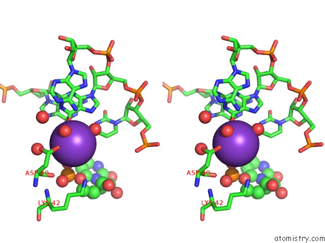
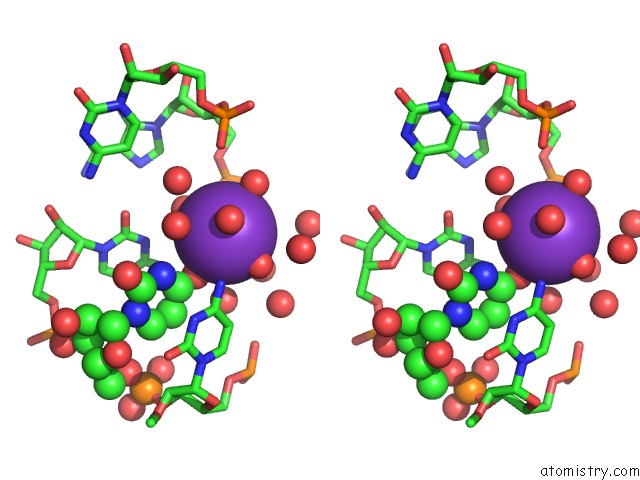
Potassium binding site 1 out of 6 in 1jbs
Go back to
Potassium binding site 1 out
of 6 in the Crystal Structure of Ribotoxin Restrictocin and A 29-Mer Srd Rna Analog
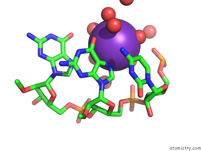
Mono view
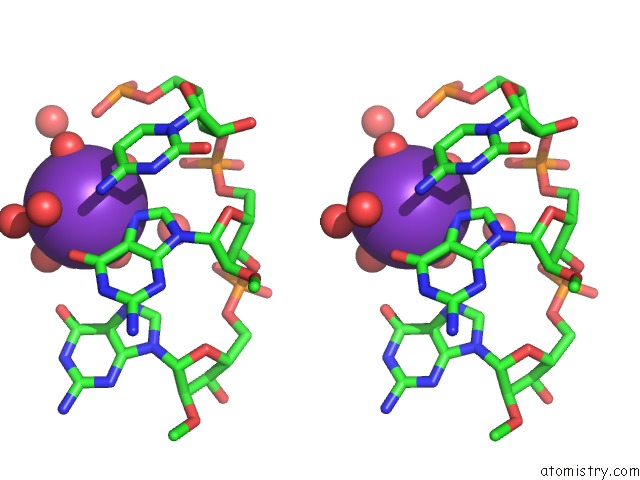
Stereo pair view
Mono view
Stereo pair view
A full contact list of Potassium with other atoms in the K binding
site number 1 of Crystal Structure of Ribotoxin Restrictocin and A 29-Mer Srd Rna Analog within 5.0Å range:
|
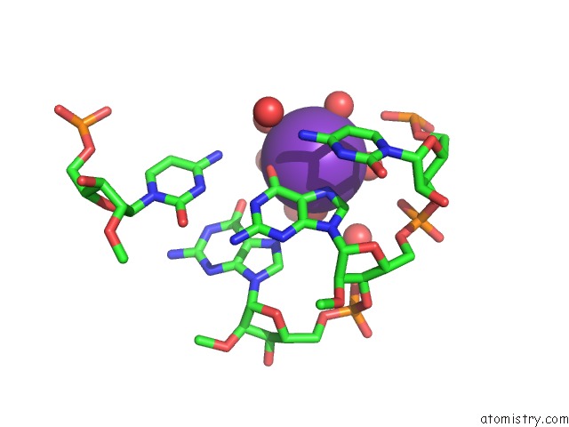
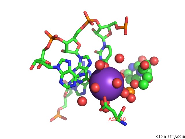
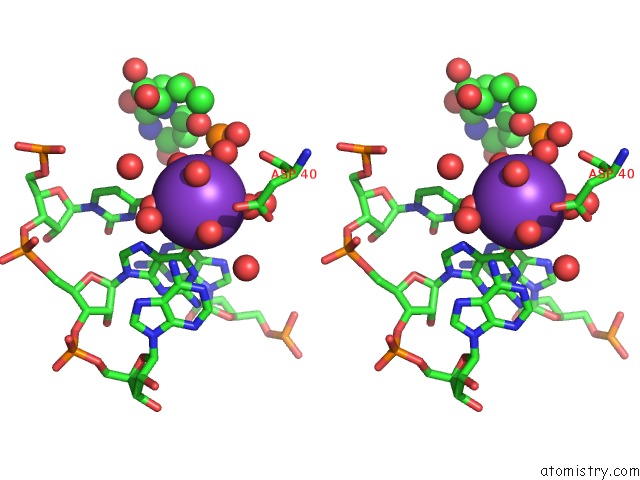
Potassium binding site 2 out of 6 in 1jbs
Go back to
Potassium binding site 2 out
of 6 in the Crystal Structure of Ribotoxin Restrictocin and A 29-Mer Srd Rna Analog
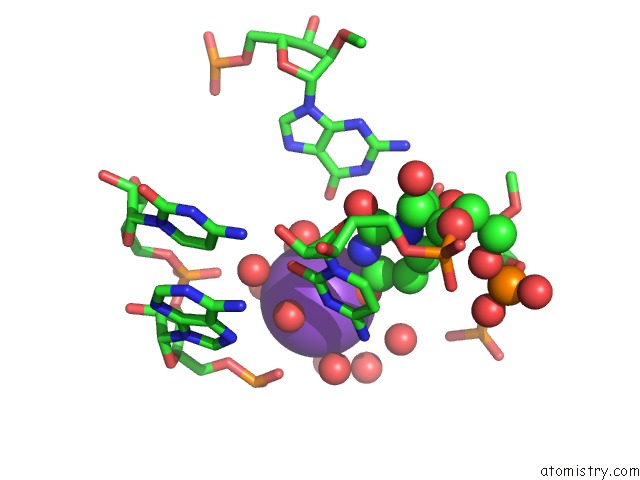
Mono view
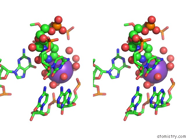
Stereo pair view
Mono view
Stereo pair view
A full contact list of Potassium with other atoms in the K binding
site number 2 of Crystal Structure of Ribotoxin Restrictocin and A 29-Mer Srd Rna Analog within 5.0Å range:
|
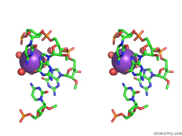
Potassium binding site 3 out of 6 in 1jbs
Go back to
Potassium binding site 3 out
of 6 in the Crystal Structure of Ribotoxin Restrictocin and A 29-Mer Srd Rna Analog

Mono view
Stereo pair view
Mono view
Stereo pair view
A full contact list of Potassium with other atoms in the K binding
site number 3 of Crystal Structure of Ribotoxin Restrictocin and A 29-Mer Srd Rna Analog within 5.0Å range:
|
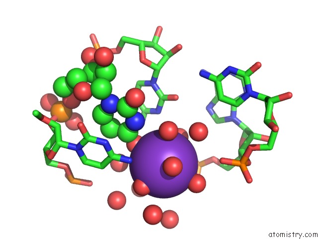
Potassium binding site 4 out of 6 in 1jbs
Go back to
Potassium binding site 4 out
of 6 in the Crystal Structure of Ribotoxin Restrictocin and A 29-Mer Srd Rna Analog
Mono view
Stereo pair view
Mono view
Stereo pair view
A full contact list of Potassium with other atoms in the K binding
site number 4 of Crystal Structure of Ribotoxin Restrictocin and A 29-Mer Srd Rna Analog within 5.0Å range:
|
Potassium binding site 5 out of 6 in 1jbs
Go back to
Potassium binding site 5 out
of 6 in the Crystal Structure of Ribotoxin Restrictocin and A 29-Mer Srd Rna Analog
Mono view
Stereo pair view
Mono view
Stereo pair view
A full contact list of Potassium with other atoms in the K binding
site number 5 of Crystal Structure of Ribotoxin Restrictocin and A 29-Mer Srd Rna Analog within 5.0Å range:
|
Potassium binding site 6 out of 6 in 1jbs
Go back to
Potassium binding site 6 out
of 6 in the Crystal Structure of Ribotoxin Restrictocin and A 29-Mer Srd Rna Analog
Mono view
Stereo pair view
Mono view
Stereo pair view
A full contact list of Potassium with other atoms in the K binding
site number 6 of Crystal Structure of Ribotoxin Restrictocin and A 29-Mer Srd Rna Analog within 5.0Å range:
|
Reference:
X.Yang,
T.Gerczei,
L.T.Glover,
C.C.Correll.
Crystal Structures of Restrictocin-Inhibitor Complexes with Implications For Rna Recognition and Base Flipping. Nat.Struct.Biol. V. 8 968 2001.
ISSN: ISSN 1072-8368
PubMed: 11685244
DOI: 10.1038/NSB1101-968
Page generated: Mon Aug 12 04:36:48 2024
ISSN: ISSN 1072-8368
PubMed: 11685244
DOI: 10.1038/NSB1101-968
Last articles
As in 2DYBAs in 2D28
As in 2BVT
As in 2BVY
As in 2BOX
As in 2BOU
As in 2BOC
As in 2BOQ
As in 1Z6B
As in 2A2T