Potassium »
PDB 1gup-1k4c »
1iwb »
Potassium in PDB 1iwb: Crystal Structure of Diol Dehydratase
Enzymatic activity of Crystal Structure of Diol Dehydratase
All present enzymatic activity of Crystal Structure of Diol Dehydratase:
4.2.1.28;
4.2.1.28;
Protein crystallography data
The structure of Crystal Structure of Diol Dehydratase, PDB code: 1iwb
was solved by
N.Shibata,
J.Masuda,
Y.Morimoto,
N.Yasuoka,
T.Toraya,
with X-Ray Crystallography technique. A brief refinement statistics is given in the table below:
| Resolution Low / High (Å) | 10.00 / 1.85 |
| Space group | P 21 21 21 |
| Cell size a, b, c (Å), α, β, γ (°) | 75.790, 122.400, 207.590, 90.00, 90.00, 90.00 |
| R / Rfree (%) | 18.1 / 25.6 |
Other elements in 1iwb:
The structure of Crystal Structure of Diol Dehydratase also contains other interesting chemical elements:
| Cobalt | (Co) | 2 atoms |
Potassium Binding Sites:
The binding sites of Potassium atom in the Crystal Structure of Diol Dehydratase
(pdb code 1iwb). This binding sites where shown within
5.0 Angstroms radius around Potassium atom.
In total 6 binding sites of Potassium where determined in the Crystal Structure of Diol Dehydratase, PDB code: 1iwb:
Jump to Potassium binding site number: 1; 2; 3; 4; 5; 6;
In total 6 binding sites of Potassium where determined in the Crystal Structure of Diol Dehydratase, PDB code: 1iwb:
Jump to Potassium binding site number: 1; 2; 3; 4; 5; 6;
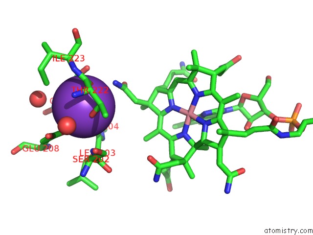
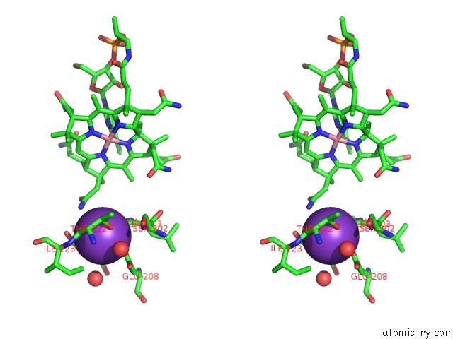
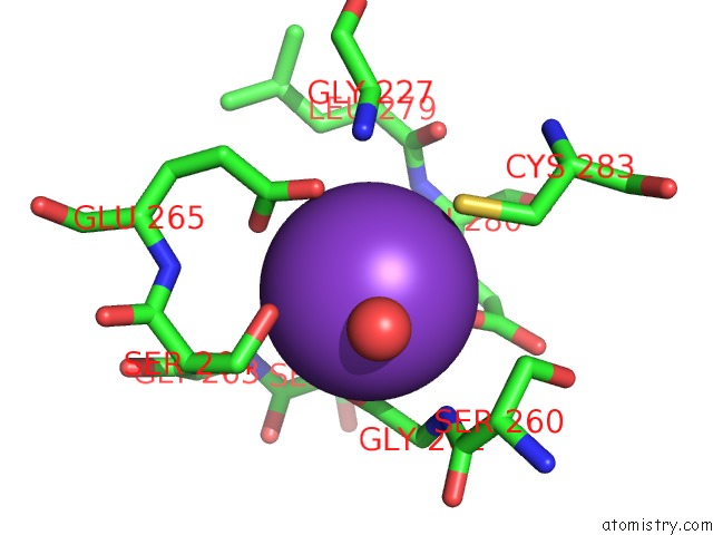
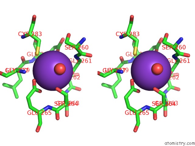
Potassium binding site 1 out of 6 in 1iwb
Go back to
Potassium binding site 1 out
of 6 in the Crystal Structure of Diol Dehydratase
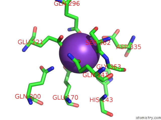
Mono view
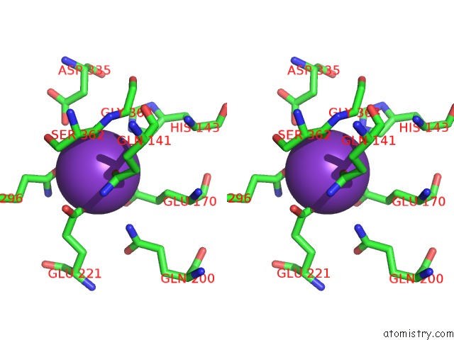
Stereo pair view
Mono view
Stereo pair view
A full contact list of Potassium with other atoms in the K binding
site number 1 of Crystal Structure of Diol Dehydratase within 5.0Å range:
|
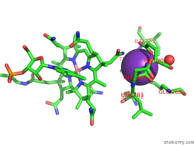
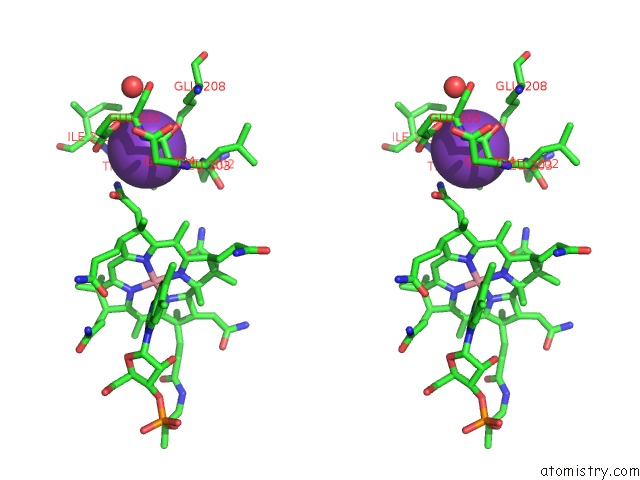
Potassium binding site 2 out of 6 in 1iwb
Go back to
Potassium binding site 2 out
of 6 in the Crystal Structure of Diol Dehydratase
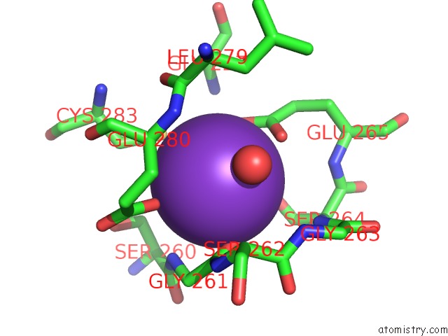
Mono view
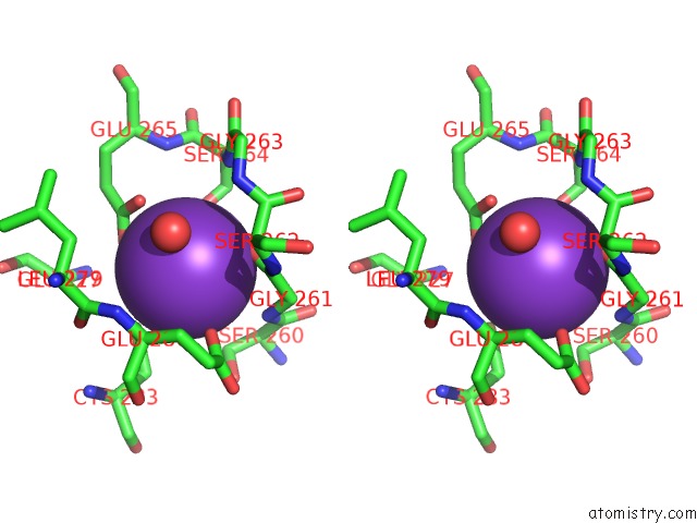
Stereo pair view
Mono view
Stereo pair view
A full contact list of Potassium with other atoms in the K binding
site number 2 of Crystal Structure of Diol Dehydratase within 5.0Å range:
|
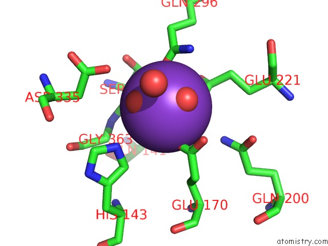
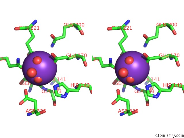
Potassium binding site 3 out of 6 in 1iwb
Go back to
Potassium binding site 3 out
of 6 in the Crystal Structure of Diol Dehydratase
Mono view
Stereo pair view
Mono view
Stereo pair view
A full contact list of Potassium with other atoms in the K binding
site number 3 of Crystal Structure of Diol Dehydratase within 5.0Å range:
|
Potassium binding site 4 out of 6 in 1iwb
Go back to
Potassium binding site 4 out
of 6 in the Crystal Structure of Diol Dehydratase
Mono view
Stereo pair view
Mono view
Stereo pair view
A full contact list of Potassium with other atoms in the K binding
site number 4 of Crystal Structure of Diol Dehydratase within 5.0Å range:
|
Potassium binding site 5 out of 6 in 1iwb
Go back to
Potassium binding site 5 out
of 6 in the Crystal Structure of Diol Dehydratase
Mono view
Stereo pair view
Mono view
Stereo pair view
A full contact list of Potassium with other atoms in the K binding
site number 5 of Crystal Structure of Diol Dehydratase within 5.0Å range:
|
Potassium binding site 6 out of 6 in 1iwb
Go back to
Potassium binding site 6 out
of 6 in the Crystal Structure of Diol Dehydratase
Mono view
Stereo pair view
Mono view
Stereo pair view
A full contact list of Potassium with other atoms in the K binding
site number 6 of Crystal Structure of Diol Dehydratase within 5.0Å range:
|
Reference:
N.Shibata,
J.Masuda,
Y.Morimoto,
N.Yasuoka,
T.Toraya.
Substrate-Induced Conformational Change of A Coenzyme B12-Dependent Enzyme: Crystal Structure of the Substrate-Free Form of Diol Dehydratase Biochemistry V. 41 12607 2002.
ISSN: ISSN 0006-2960
PubMed: 12379103
DOI: 10.1021/BI026104Z
Page generated: Mon Aug 12 04:34:33 2024
ISSN: ISSN 0006-2960
PubMed: 12379103
DOI: 10.1021/BI026104Z
Last articles
Zn in 9J0NZn in 9J0O
Zn in 9J0P
Zn in 9FJX
Zn in 9EKB
Zn in 9C0F
Zn in 9CAH
Zn in 9CH0
Zn in 9CH3
Zn in 9CH1