Potassium »
PDB 1gup-1k4c »
1h54 »
Potassium in PDB 1h54: Maltose Phosphorylase From Lactobacillus Brevis
Enzymatic activity of Maltose Phosphorylase From Lactobacillus Brevis
All present enzymatic activity of Maltose Phosphorylase From Lactobacillus Brevis:
2.4.1.8;
2.4.1.8;
Protein crystallography data
The structure of Maltose Phosphorylase From Lactobacillus Brevis, PDB code: 1h54
was solved by
H.Van Tilbeurgh,
M.-P.Egloff,
with X-Ray Crystallography technique. A brief refinement statistics is given in the table below:
| Resolution Low / High (Å) | 29.1 / 2.15 |
| Space group | P 1 21 1 |
| Cell size a, b, c (Å), α, β, γ (°) | 83.750, 102.500, 114.000, 90.00, 111.30, 90.00 |
| R / Rfree (%) | 18.6 / 22.5 |
Potassium Binding Sites:
The binding sites of Potassium atom in the Maltose Phosphorylase From Lactobacillus Brevis
(pdb code 1h54). This binding sites where shown within
5.0 Angstroms radius around Potassium atom.
In total 2 binding sites of Potassium where determined in the Maltose Phosphorylase From Lactobacillus Brevis, PDB code: 1h54:
Jump to Potassium binding site number: 1; 2;
In total 2 binding sites of Potassium where determined in the Maltose Phosphorylase From Lactobacillus Brevis, PDB code: 1h54:
Jump to Potassium binding site number: 1; 2;
Potassium binding site 1 out of 2 in 1h54
Go back to
Potassium binding site 1 out
of 2 in the Maltose Phosphorylase From Lactobacillus Brevis
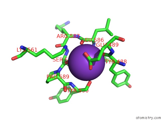
Mono view
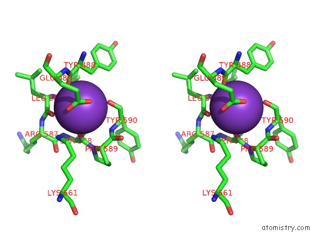
Stereo pair view
Mono view
Stereo pair view
A full contact list of Potassium with other atoms in the K binding
site number 1 of Maltose Phosphorylase From Lactobacillus Brevis within 5.0Å range:
|
Potassium binding site 2 out of 2 in 1h54
Go back to
Potassium binding site 2 out
of 2 in the Maltose Phosphorylase From Lactobacillus Brevis
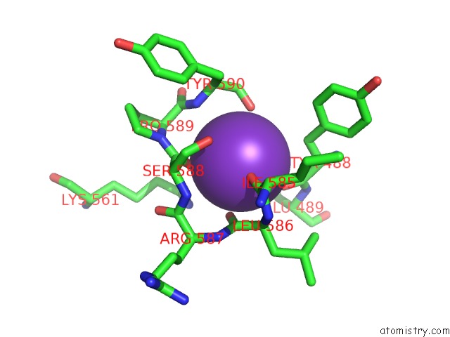
Mono view
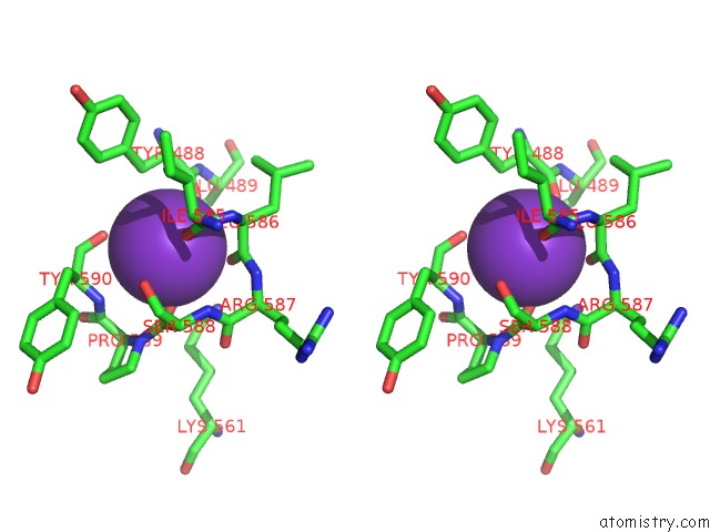
Stereo pair view
Mono view
Stereo pair view
A full contact list of Potassium with other atoms in the K binding
site number 2 of Maltose Phosphorylase From Lactobacillus Brevis within 5.0Å range:
|
Reference:
M.-P.Egloff,
J.Uppenberg,
L.Haalck,
H.Van Tilbeurgh.
Crystal Structure of Maltose Phosphorylase From Lactobacillus Brevis: Unexpected Evolutionary Relationship with Glucoamylases. Structure V. 9 689 2001.
ISSN: ISSN 0969-2126
PubMed: 11587643
DOI: 10.1016/S0969-2126(01)00626-8
Page generated: Mon Aug 12 04:32:22 2024
ISSN: ISSN 0969-2126
PubMed: 11587643
DOI: 10.1016/S0969-2126(01)00626-8
Last articles
Zn in 9JYWZn in 9IR4
Zn in 9IR3
Zn in 9GMX
Zn in 9GMW
Zn in 9JEJ
Zn in 9ERF
Zn in 9ERE
Zn in 9EGV
Zn in 9EGW