Potassium »
PDB 1d7v-1gmk »
1g8m »
Potassium in PDB 1g8m: Crystal Structure of Avian Atic, A Bifunctional Transformylase and Cyclohydrolase Enzyme in Purine Biosynthesis at 1.75 Ang. Resolution
Enzymatic activity of Crystal Structure of Avian Atic, A Bifunctional Transformylase and Cyclohydrolase Enzyme in Purine Biosynthesis at 1.75 Ang. Resolution
All present enzymatic activity of Crystal Structure of Avian Atic, A Bifunctional Transformylase and Cyclohydrolase Enzyme in Purine Biosynthesis at 1.75 Ang. Resolution:
2.1.2.3; 3.5.4.10;
2.1.2.3; 3.5.4.10;
Protein crystallography data
The structure of Crystal Structure of Avian Atic, A Bifunctional Transformylase and Cyclohydrolase Enzyme in Purine Biosynthesis at 1.75 Ang. Resolution, PDB code: 1g8m
was solved by
S.E.Greasley,
P.Horton,
G.P.Beardsley,
S.J.Benkovic,
I.A.Wilson,
with X-Ray Crystallography technique. A brief refinement statistics is given in the table below:
| Resolution Low / High (Å) | 50.00 / 1.75 |
| Space group | P 1 21 1 |
| Cell size a, b, c (Å), α, β, γ (°) | 65.100, 106.000, 103.500, 90.00, 108.00, 90.00 |
| R / Rfree (%) | 20 / 21.6 |
Potassium Binding Sites:
The binding sites of Potassium atom in the Crystal Structure of Avian Atic, A Bifunctional Transformylase and Cyclohydrolase Enzyme in Purine Biosynthesis at 1.75 Ang. Resolution
(pdb code 1g8m). This binding sites where shown within
5.0 Angstroms radius around Potassium atom.
In total 2 binding sites of Potassium where determined in the Crystal Structure of Avian Atic, A Bifunctional Transformylase and Cyclohydrolase Enzyme in Purine Biosynthesis at 1.75 Ang. Resolution, PDB code: 1g8m:
Jump to Potassium binding site number: 1; 2;
In total 2 binding sites of Potassium where determined in the Crystal Structure of Avian Atic, A Bifunctional Transformylase and Cyclohydrolase Enzyme in Purine Biosynthesis at 1.75 Ang. Resolution, PDB code: 1g8m:
Jump to Potassium binding site number: 1; 2;
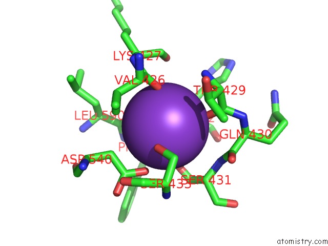
Potassium binding site 1 out of 2 in 1g8m
Go back to
Potassium binding site 1 out
of 2 in the Crystal Structure of Avian Atic, A Bifunctional Transformylase and Cyclohydrolase Enzyme in Purine Biosynthesis at 1.75 Ang. Resolution

Mono view

Stereo pair view
Mono view
Stereo pair view
A full contact list of Potassium with other atoms in the K binding
site number 1 of Crystal Structure of Avian Atic, A Bifunctional Transformylase and Cyclohydrolase Enzyme in Purine Biosynthesis at 1.75 Ang. Resolution within 5.0Å range:
|
Potassium binding site 2 out of 2 in 1g8m
Go back to
Potassium binding site 2 out
of 2 in the Crystal Structure of Avian Atic, A Bifunctional Transformylase and Cyclohydrolase Enzyme in Purine Biosynthesis at 1.75 Ang. Resolution
Mono view

Stereo pair view
Mono view
Stereo pair view
A full contact list of Potassium with other atoms in the K binding
site number 2 of Crystal Structure of Avian Atic, A Bifunctional Transformylase and Cyclohydrolase Enzyme in Purine Biosynthesis at 1.75 Ang. Resolution within 5.0Å range:
|
Reference:
S.E.Greasley,
P.Horton,
J.Ramcharan,
G.P.Beardsley,
S.J.Benkovic,
I.A.Wilson.
Crystal Structure of A Bifunctional Transformylase and Cyclohydrolase Enzyme in Purine Biosynthesis. Nat.Struct.Biol. V. 8 402 2001.
ISSN: ISSN 1072-8368
PubMed: 11323713
DOI: 10.1038/87555
Page generated: Mon Aug 12 04:30:03 2024
ISSN: ISSN 1072-8368
PubMed: 11323713
DOI: 10.1038/87555
Last articles
Zn in 9J0NZn in 9J0O
Zn in 9J0P
Zn in 9FJX
Zn in 9EKB
Zn in 9C0F
Zn in 9CAH
Zn in 9CH0
Zn in 9CH3
Zn in 9CH1