Potassium »
PDB 1d7v-1gmk »
1f7y »
Potassium in PDB 1f7y: The Crystal Structure of Two Uucg Loops Highlights the Role Played By 2'-Hydroxyl Groups in Its Unusual Stability
Protein crystallography data
The structure of The Crystal Structure of Two Uucg Loops Highlights the Role Played By 2'-Hydroxyl Groups in Its Unusual Stability, PDB code: 1f7y
was solved by
E.Ennifar,
A.Nikouline,
A.Serganov,
S.Tishchenko,
N.Nevskaya,
M.Garber,
B.Ehresmann,
C.Ehresmann,
S.Nikonov,
P.Dumas,
with X-Ray Crystallography technique. A brief refinement statistics is given in the table below:
| Resolution Low / High (Å) | 8.00 / 2.80 |
| Space group | P 64 2 2 |
| Cell size a, b, c (Å), α, β, γ (°) | 128.800, 128.800, 65.100, 90.00, 90.00, 120.00 |
| R / Rfree (%) | 22.2 / 28.9 |
Other elements in 1f7y:
The structure of The Crystal Structure of Two Uucg Loops Highlights the Role Played By 2'-Hydroxyl Groups in Its Unusual Stability also contains other interesting chemical elements:
| Magnesium | (Mg) | 9 atoms |
| Sodium | (Na) | 2 atoms |
Potassium Binding Sites:
The binding sites of Potassium atom in the The Crystal Structure of Two Uucg Loops Highlights the Role Played By 2'-Hydroxyl Groups in Its Unusual Stability
(pdb code 1f7y). This binding sites where shown within
5.0 Angstroms radius around Potassium atom.
In total only one binding site of Potassium was determined in the The Crystal Structure of Two Uucg Loops Highlights the Role Played By 2'-Hydroxyl Groups in Its Unusual Stability, PDB code: 1f7y:
In total only one binding site of Potassium was determined in the The Crystal Structure of Two Uucg Loops Highlights the Role Played By 2'-Hydroxyl Groups in Its Unusual Stability, PDB code: 1f7y:
Potassium binding site 1 out of 1 in 1f7y
Go back to
Potassium binding site 1 out
of 1 in the The Crystal Structure of Two Uucg Loops Highlights the Role Played By 2'-Hydroxyl Groups in Its Unusual Stability
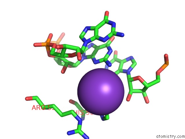
Mono view
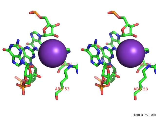
Stereo pair view
Mono view
Stereo pair view
A full contact list of Potassium with other atoms in the K binding
site number 1 of The Crystal Structure of Two Uucg Loops Highlights the Role Played By 2'-Hydroxyl Groups in Its Unusual Stability within 5.0Å range:
|
Reference:
E.Ennifar,
A.Nikulin,
S.Tishchenko,
A.Serganov,
N.Nevskaya,
M.Garber,
B.Ehresmann,
C.Ehresmann,
S.Nikonov,
P.Dumas.
The Crystal Structure of Uucg Tetraloop. J.Mol.Biol. V. 304 35 2000.
ISSN: ISSN 0022-2836
PubMed: 11071808
DOI: 10.1006/JMBI.2000.4204
Page generated: Mon Aug 12 04:28:11 2024
ISSN: ISSN 0022-2836
PubMed: 11071808
DOI: 10.1006/JMBI.2000.4204
Last articles
Zn in 9J0NZn in 9J0O
Zn in 9J0P
Zn in 9FJX
Zn in 9EKB
Zn in 9C0F
Zn in 9CAH
Zn in 9CH0
Zn in 9CH3
Zn in 9CH1