Potassium »
PDB 1d7v-1gmk »
1dul »
Potassium in PDB 1dul: Structure of the Ribonucleoprotein Core of the E. Coli Signal Recognition Particle
Protein crystallography data
The structure of Structure of the Ribonucleoprotein Core of the E. Coli Signal Recognition Particle, PDB code: 1dul
was solved by
R.T.Batey,
R.P.Rambo,
L.Lucast,
B.Rha,
J.Doudna,
with X-Ray Crystallography technique. A brief refinement statistics is given in the table below:
| Resolution Low / High (Å) | 28.83 / 1.80 |
| Space group | C 1 2 1 |
| Cell size a, b, c (Å), α, β, γ (°) | 136.567, 78.291, 32.849, 90.00, 96.14, 90.00 |
| R / Rfree (%) | 19.9 / 22.1 |
Other elements in 1dul:
The structure of Structure of the Ribonucleoprotein Core of the E. Coli Signal Recognition Particle also contains other interesting chemical elements:
| Magnesium | (Mg) | 4 atoms |
Potassium Binding Sites:
The binding sites of Potassium atom in the Structure of the Ribonucleoprotein Core of the E. Coli Signal Recognition Particle
(pdb code 1dul). This binding sites where shown within
5.0 Angstroms radius around Potassium atom.
In total 3 binding sites of Potassium where determined in the Structure of the Ribonucleoprotein Core of the E. Coli Signal Recognition Particle, PDB code: 1dul:
Jump to Potassium binding site number: 1; 2; 3;
In total 3 binding sites of Potassium where determined in the Structure of the Ribonucleoprotein Core of the E. Coli Signal Recognition Particle, PDB code: 1dul:
Jump to Potassium binding site number: 1; 2; 3;
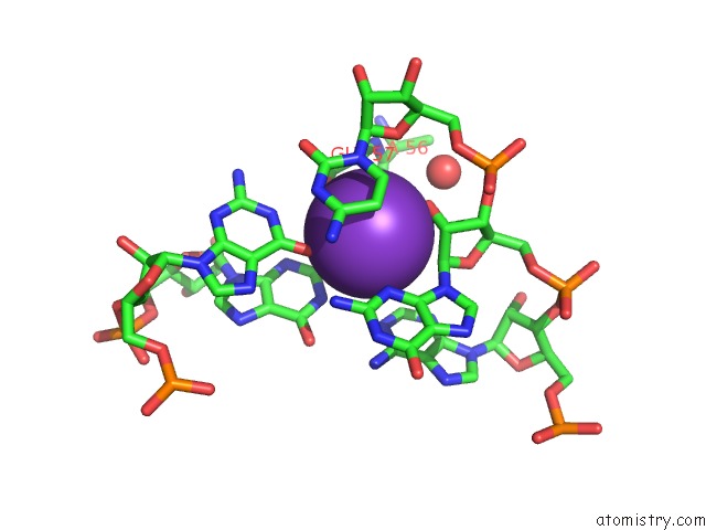
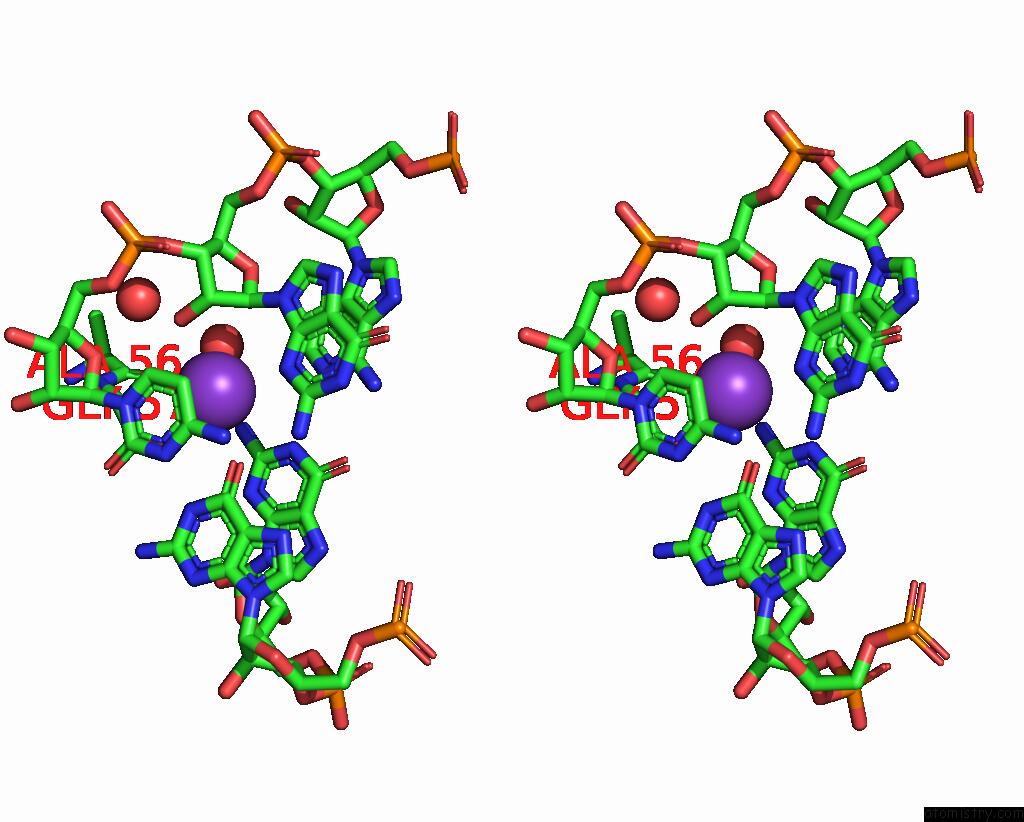
Potassium binding site 1 out of 3 in 1dul
Go back to
Potassium binding site 1 out
of 3 in the Structure of the Ribonucleoprotein Core of the E. Coli Signal Recognition Particle
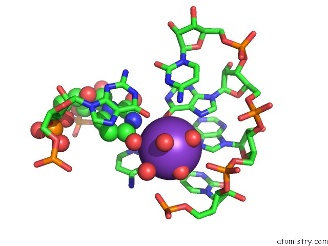
Mono view
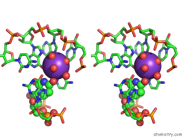
Stereo pair view
Mono view
Stereo pair view
A full contact list of Potassium with other atoms in the K binding
site number 1 of Structure of the Ribonucleoprotein Core of the E. Coli Signal Recognition Particle within 5.0Å range:
|
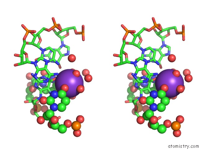
Potassium binding site 2 out of 3 in 1dul
Go back to
Potassium binding site 2 out
of 3 in the Structure of the Ribonucleoprotein Core of the E. Coli Signal Recognition Particle
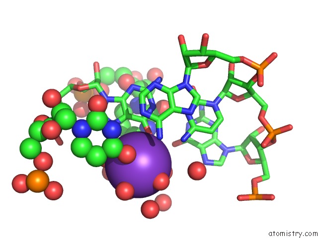
Mono view
Stereo pair view
Mono view
Stereo pair view
A full contact list of Potassium with other atoms in the K binding
site number 2 of Structure of the Ribonucleoprotein Core of the E. Coli Signal Recognition Particle within 5.0Å range:
|
Potassium binding site 3 out of 3 in 1dul
Go back to
Potassium binding site 3 out
of 3 in the Structure of the Ribonucleoprotein Core of the E. Coli Signal Recognition Particle
Mono view
Stereo pair view
Mono view
Stereo pair view
A full contact list of Potassium with other atoms in the K binding
site number 3 of Structure of the Ribonucleoprotein Core of the E. Coli Signal Recognition Particle within 5.0Å range:
|
Reference:
R.T.Batey,
R.P.Rambo,
L.Lucast,
B.Rha,
J.A.Doudna.
Crystal Structure of the Ribonucleoprotein Core of the Signal Recognition Particle. Science V. 287 1232 2000.
ISSN: ISSN 0036-8075
PubMed: 10678824
DOI: 10.1126/SCIENCE.287.5456.1232
Page generated: Mon Aug 12 04:24:21 2024
ISSN: ISSN 0036-8075
PubMed: 10678824
DOI: 10.1126/SCIENCE.287.5456.1232
Last articles
Zn in 9MJ5Zn in 9HNW
Zn in 9G0L
Zn in 9FNE
Zn in 9DZN
Zn in 9E0I
Zn in 9D32
Zn in 9DAK
Zn in 8ZXC
Zn in 8ZUF