Potassium »
PDB 1a3w-1d7u »
1d7r »
Potassium in PDB 1d7r: Crystal Structure of the Complex of 2,2-Dialkylglycine Decarboxylase with 5PA
Enzymatic activity of Crystal Structure of the Complex of 2,2-Dialkylglycine Decarboxylase with 5PA
All present enzymatic activity of Crystal Structure of the Complex of 2,2-Dialkylglycine Decarboxylase with 5PA:
4.1.1.64;
4.1.1.64;
Protein crystallography data
The structure of Crystal Structure of the Complex of 2,2-Dialkylglycine Decarboxylase with 5PA, PDB code: 1d7r
was solved by
V.N.Malashkevich,
M.D.Toney,
P.Strop,
J.Keller,
J.N.Jansonius,
with X-Ray Crystallography technique. A brief refinement statistics is given in the table below:
| Resolution Low / High (Å) | 10.00 / 2.00 |
| Space group | P 64 2 2 |
| Cell size a, b, c (Å), α, β, γ (°) | 152.650, 152.650, 86.220, 90.00, 90.00, 120.00 |
| R / Rfree (%) | n/a / n/a |
Other elements in 1d7r:
The structure of Crystal Structure of the Complex of 2,2-Dialkylglycine Decarboxylase with 5PA also contains other interesting chemical elements:
| Sodium | (Na) | 1 atom |
Potassium Binding Sites:
The binding sites of Potassium atom in the Crystal Structure of the Complex of 2,2-Dialkylglycine Decarboxylase with 5PA
(pdb code 1d7r). This binding sites where shown within
5.0 Angstroms radius around Potassium atom.
In total only one binding site of Potassium was determined in the Crystal Structure of the Complex of 2,2-Dialkylglycine Decarboxylase with 5PA, PDB code: 1d7r:
In total only one binding site of Potassium was determined in the Crystal Structure of the Complex of 2,2-Dialkylglycine Decarboxylase with 5PA, PDB code: 1d7r:
Potassium binding site 1 out of 1 in 1d7r
Go back to
Potassium binding site 1 out
of 1 in the Crystal Structure of the Complex of 2,2-Dialkylglycine Decarboxylase with 5PA
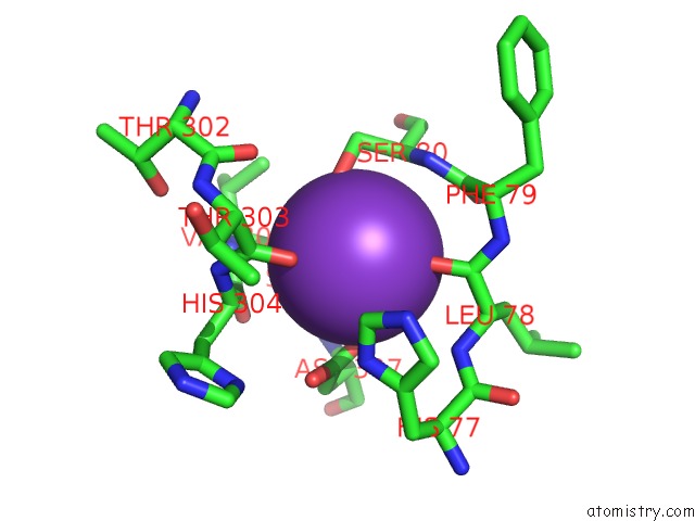
Mono view
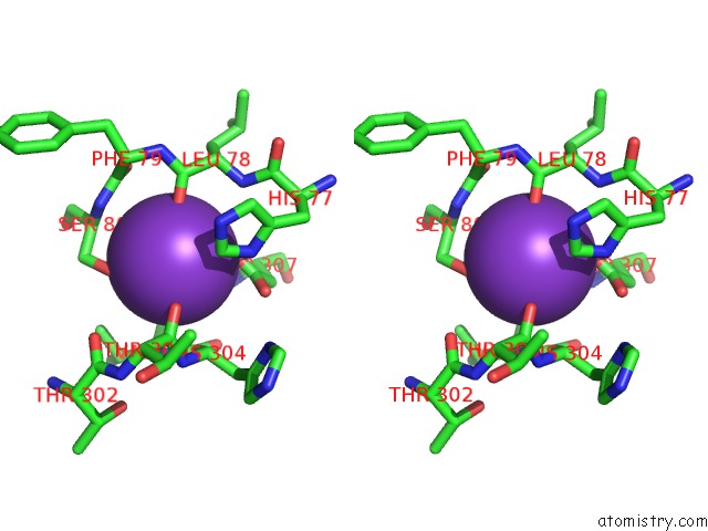
Stereo pair view
Mono view
Stereo pair view
A full contact list of Potassium with other atoms in the K binding
site number 1 of Crystal Structure of the Complex of 2,2-Dialkylglycine Decarboxylase with 5PA within 5.0Å range:
|
Reference:
V.N.Malashkevich,
P.Strop,
J.W.Keller,
J.N.Jansonius,
M.D.Toney.
Crystal Structures of Dialkylglycine Decarboxylase Inhibitor Complexes. J.Mol.Biol. V. 294 193 1999.
ISSN: ISSN 0022-2836
PubMed: 10556038
DOI: 10.1006/JMBI.1999.3254
Page generated: Mon Aug 12 04:18:31 2024
ISSN: ISSN 0022-2836
PubMed: 10556038
DOI: 10.1006/JMBI.1999.3254
Last articles
Zn in 9J0NZn in 9J0O
Zn in 9J0P
Zn in 9FJX
Zn in 9EKB
Zn in 9C0F
Zn in 9CAH
Zn in 9CH0
Zn in 9CH3
Zn in 9CH1