Potassium »
PDB 1a3w-1d7u »
1bw9 »
Potassium in PDB 1bw9: Phenylalanine Dehydrogenase Structure in Ternary Complex with Nad+ and Phenylpyruvate
Enzymatic activity of Phenylalanine Dehydrogenase Structure in Ternary Complex with Nad+ and Phenylpyruvate
All present enzymatic activity of Phenylalanine Dehydrogenase Structure in Ternary Complex with Nad+ and Phenylpyruvate:
1.4.1.20;
1.4.1.20;
Protein crystallography data
The structure of Phenylalanine Dehydrogenase Structure in Ternary Complex with Nad+ and Phenylpyruvate, PDB code: 1bw9
was solved by
J.L.Vanhooke,
J.B.Thoden,
N.M.W.Brunhuber,
J.L.Blanchard,
H.M.Holden,
with X-Ray Crystallography technique. A brief refinement statistics is given in the table below:
| Resolution Low / High (Å) | 30.00 / 1.50 |
| Space group | P 21 21 21 |
| Cell size a, b, c (Å), α, β, γ (°) | 64.300, 110.200, 113.400, 90.00, 90.00, 90.00 |
| R / Rfree (%) | n/a / n/a |
Other elements in 1bw9:
The structure of Phenylalanine Dehydrogenase Structure in Ternary Complex with Nad+ and Phenylpyruvate also contains other interesting chemical elements:
| Sodium | (Na) | 2 atoms |
Potassium Binding Sites:
The binding sites of Potassium atom in the Phenylalanine Dehydrogenase Structure in Ternary Complex with Nad+ and Phenylpyruvate
(pdb code 1bw9). This binding sites where shown within
5.0 Angstroms radius around Potassium atom.
In total 2 binding sites of Potassium where determined in the Phenylalanine Dehydrogenase Structure in Ternary Complex with Nad+ and Phenylpyruvate, PDB code: 1bw9:
Jump to Potassium binding site number: 1; 2;
In total 2 binding sites of Potassium where determined in the Phenylalanine Dehydrogenase Structure in Ternary Complex with Nad+ and Phenylpyruvate, PDB code: 1bw9:
Jump to Potassium binding site number: 1; 2;
Potassium binding site 1 out of 2 in 1bw9
Go back to
Potassium binding site 1 out
of 2 in the Phenylalanine Dehydrogenase Structure in Ternary Complex with Nad+ and Phenylpyruvate
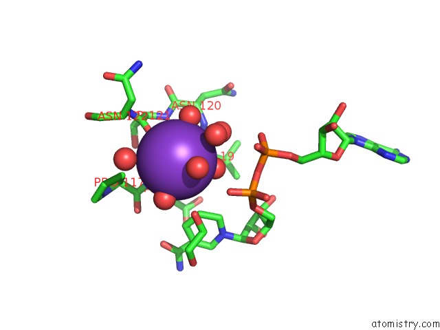
Mono view
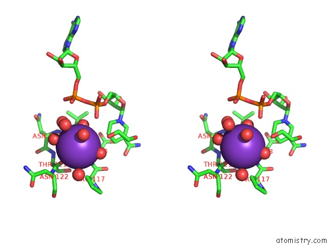
Stereo pair view
Mono view
Stereo pair view
A full contact list of Potassium with other atoms in the K binding
site number 1 of Phenylalanine Dehydrogenase Structure in Ternary Complex with Nad+ and Phenylpyruvate within 5.0Å range:
|
Potassium binding site 2 out of 2 in 1bw9
Go back to
Potassium binding site 2 out
of 2 in the Phenylalanine Dehydrogenase Structure in Ternary Complex with Nad+ and Phenylpyruvate
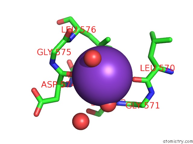
Mono view
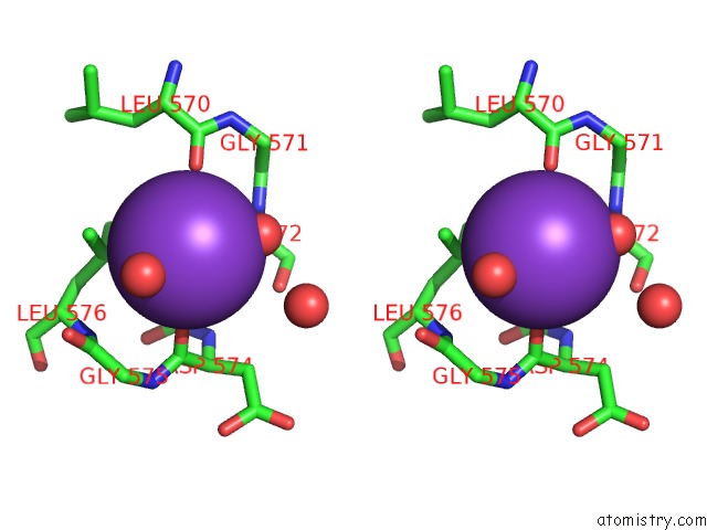
Stereo pair view
Mono view
Stereo pair view
A full contact list of Potassium with other atoms in the K binding
site number 2 of Phenylalanine Dehydrogenase Structure in Ternary Complex with Nad+ and Phenylpyruvate within 5.0Å range:
|
Reference:
J.L.Vanhooke,
J.B.Thoden,
N.M.Brunhuber,
J.S.Blanchard,
H.M.Holden.
Phenylalanine Dehydrogenase From Rhodococcus Sp. M4: High-Resolution X-Ray Analyses of Inhibitory Ternary Complexes Reveal Key Features in the Oxidative Deamination Mechanism. Biochemistry V. 38 2326 1999.
ISSN: ISSN 0006-2960
PubMed: 10029526
DOI: 10.1021/BI982244Q
Page generated: Mon Aug 12 04:04:54 2024
ISSN: ISSN 0006-2960
PubMed: 10029526
DOI: 10.1021/BI982244Q
Last articles
Zn in 9J0NZn in 9J0O
Zn in 9J0P
Zn in 9FJX
Zn in 9EKB
Zn in 9C0F
Zn in 9CAH
Zn in 9CH0
Zn in 9CH3
Zn in 9CH1