Potassium »
PDB 1a3w-1d7u »
1apx »
Potassium in PDB 1apx: Crystal Structure of Recombinant Ascorbate Peroxidase
Enzymatic activity of Crystal Structure of Recombinant Ascorbate Peroxidase
All present enzymatic activity of Crystal Structure of Recombinant Ascorbate Peroxidase:
1.11.1.11;
1.11.1.11;
Protein crystallography data
The structure of Crystal Structure of Recombinant Ascorbate Peroxidase, PDB code: 1apx
was solved by
W.R.Patterson,
T.L.Poulos,
with X-Ray Crystallography technique. A brief refinement statistics is given in the table below:
| Resolution Low / High (Å) | 8.00 / 2.20 |
| Space group | C 1 2 1 |
| Cell size a, b, c (Å), α, β, γ (°) | 132.400, 53.000, 170.600, 90.00, 107.10, 90.00 |
| R / Rfree (%) | 19.3 / n/a |
Other elements in 1apx:
The structure of Crystal Structure of Recombinant Ascorbate Peroxidase also contains other interesting chemical elements:
| Iron | (Fe) | 4 atoms |
Potassium Binding Sites:
The binding sites of Potassium atom in the Crystal Structure of Recombinant Ascorbate Peroxidase
(pdb code 1apx). This binding sites where shown within
5.0 Angstroms radius around Potassium atom.
In total 4 binding sites of Potassium where determined in the Crystal Structure of Recombinant Ascorbate Peroxidase, PDB code: 1apx:
Jump to Potassium binding site number: 1; 2; 3; 4;
In total 4 binding sites of Potassium where determined in the Crystal Structure of Recombinant Ascorbate Peroxidase, PDB code: 1apx:
Jump to Potassium binding site number: 1; 2; 3; 4;
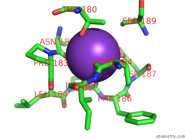
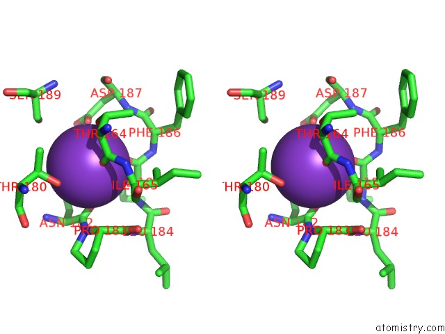
Potassium binding site 1 out of 4 in 1apx
Go back to
Potassium binding site 1 out
of 4 in the Crystal Structure of Recombinant Ascorbate Peroxidase
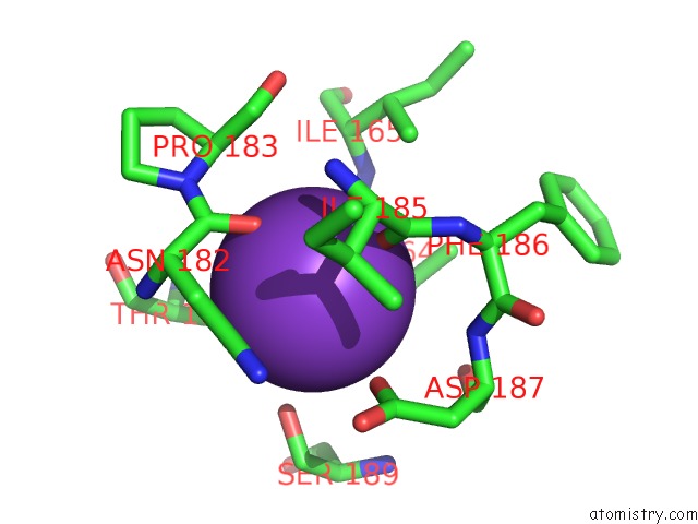
Mono view
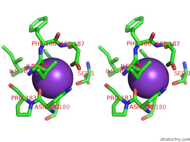
Stereo pair view
Mono view
Stereo pair view
A full contact list of Potassium with other atoms in the K binding
site number 1 of Crystal Structure of Recombinant Ascorbate Peroxidase within 5.0Å range:
|
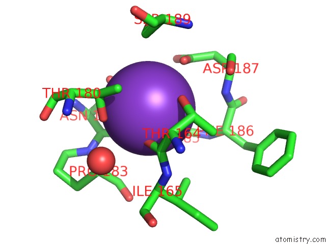
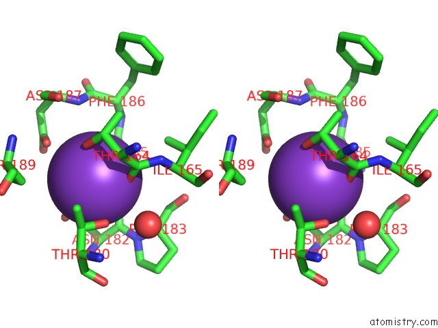
Potassium binding site 2 out of 4 in 1apx
Go back to
Potassium binding site 2 out
of 4 in the Crystal Structure of Recombinant Ascorbate Peroxidase
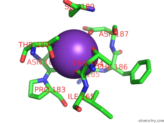
Mono view
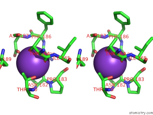
Stereo pair view
Mono view
Stereo pair view
A full contact list of Potassium with other atoms in the K binding
site number 2 of Crystal Structure of Recombinant Ascorbate Peroxidase within 5.0Å range:
|
Potassium binding site 3 out of 4 in 1apx
Go back to
Potassium binding site 3 out
of 4 in the Crystal Structure of Recombinant Ascorbate Peroxidase
Mono view
Stereo pair view
Mono view
Stereo pair view
A full contact list of Potassium with other atoms in the K binding
site number 3 of Crystal Structure of Recombinant Ascorbate Peroxidase within 5.0Å range:
|
Potassium binding site 4 out of 4 in 1apx
Go back to
Potassium binding site 4 out
of 4 in the Crystal Structure of Recombinant Ascorbate Peroxidase
Mono view
Stereo pair view
Mono view
Stereo pair view
A full contact list of Potassium with other atoms in the K binding
site number 4 of Crystal Structure of Recombinant Ascorbate Peroxidase within 5.0Å range:
|
Reference:
W.R.Patterson,
T.L.Poulos.
Crystal Structure of Recombinant Pea Cytosolic Ascorbate Peroxidase. Biochemistry V. 34 4331 1995.
ISSN: ISSN 0006-2960
PubMed: 7703247
DOI: 10.1021/BI00013A023
Page generated: Mon Aug 12 04:01:59 2024
ISSN: ISSN 0006-2960
PubMed: 7703247
DOI: 10.1021/BI00013A023
Last articles
Zn in 9J0NZn in 9J0O
Zn in 9J0P
Zn in 9FJX
Zn in 9EKB
Zn in 9C0F
Zn in 9CAH
Zn in 9CH0
Zn in 9CH3
Zn in 9CH1