Potassium »
PDB 1a3w-1d7u »
1akd »
Potassium in PDB 1akd: Cytochrome P450CAM From Pseudomonas Putida, Complexed with 1S-Camphor
Enzymatic activity of Cytochrome P450CAM From Pseudomonas Putida, Complexed with 1S-Camphor
All present enzymatic activity of Cytochrome P450CAM From Pseudomonas Putida, Complexed with 1S-Camphor:
1.14.15.1;
1.14.15.1;
Protein crystallography data
The structure of Cytochrome P450CAM From Pseudomonas Putida, Complexed with 1S-Camphor, PDB code: 1akd
was solved by
I.Schlichting,
C.Jung,
H.Schulze,
with X-Ray Crystallography technique. A brief refinement statistics is given in the table below:
| Resolution Low / High (Å) | 26.00 / 1.80 |
| Space group | P 21 21 21 |
| Cell size a, b, c (Å), α, β, γ (°) | 64.300, 66.200, 106.800, 90.00, 90.00, 90.00 |
| R / Rfree (%) | 20.9 / 26.5 |
Other elements in 1akd:
The structure of Cytochrome P450CAM From Pseudomonas Putida, Complexed with 1S-Camphor also contains other interesting chemical elements:
| Iron | (Fe) | 1 atom |
Potassium Binding Sites:
The binding sites of Potassium atom in the Cytochrome P450CAM From Pseudomonas Putida, Complexed with 1S-Camphor
(pdb code 1akd). This binding sites where shown within
5.0 Angstroms radius around Potassium atom.
In total only one binding site of Potassium was determined in the Cytochrome P450CAM From Pseudomonas Putida, Complexed with 1S-Camphor, PDB code: 1akd:
In total only one binding site of Potassium was determined in the Cytochrome P450CAM From Pseudomonas Putida, Complexed with 1S-Camphor, PDB code: 1akd:
Potassium binding site 1 out of 1 in 1akd
Go back to
Potassium binding site 1 out
of 1 in the Cytochrome P450CAM From Pseudomonas Putida, Complexed with 1S-Camphor
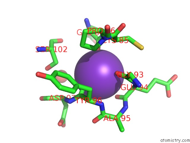
Mono view
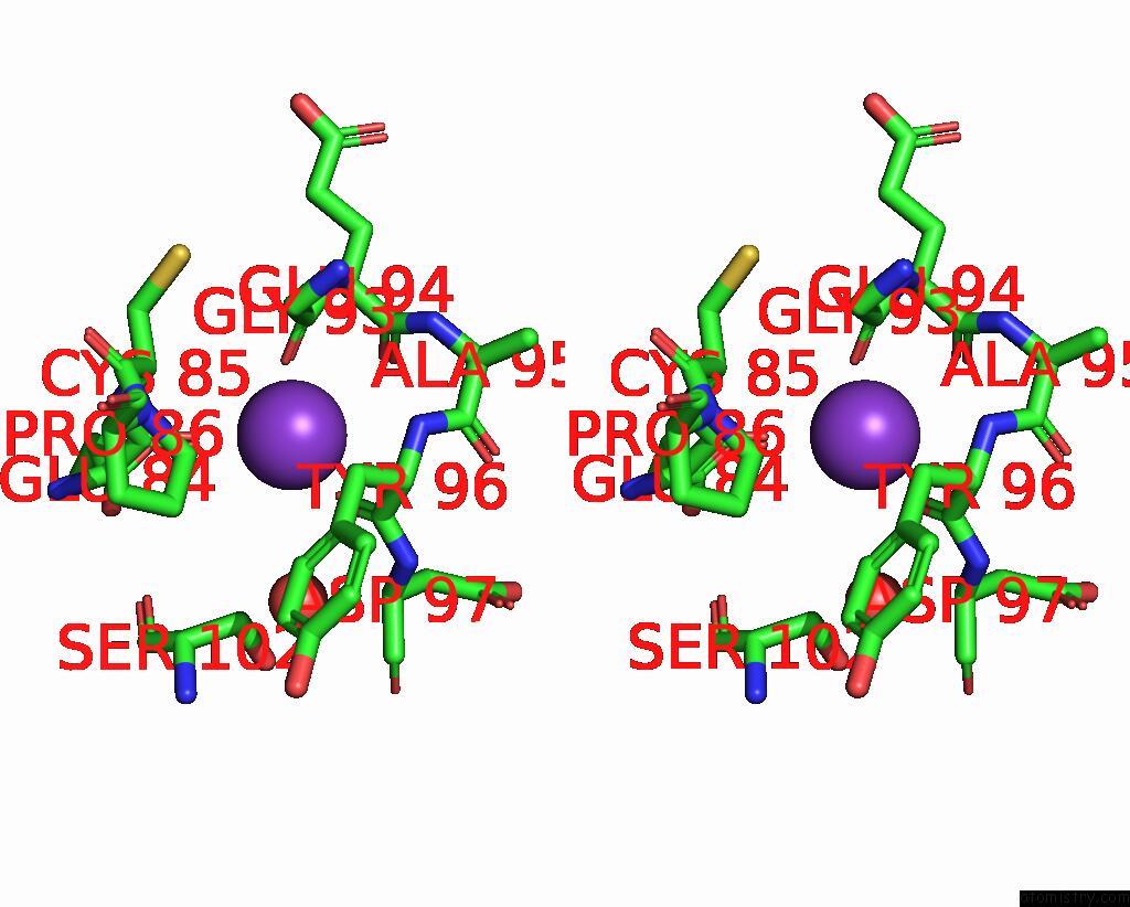
Stereo pair view
Mono view
Stereo pair view
A full contact list of Potassium with other atoms in the K binding
site number 1 of Cytochrome P450CAM From Pseudomonas Putida, Complexed with 1S-Camphor within 5.0Å range:
|
Reference:
I.Schlichting,
C.Jung,
H.Schulze.
Crystal Structure of Cytochrome P-450CAM Complexed with the (1S)-Camphor Enantiomer. Febs Lett. V. 415 253 1997.
ISSN: ISSN 0014-5793
PubMed: 9357977
DOI: 10.1016/S0014-5793(97)01135-6
Page generated: Sat Aug 9 01:32:17 2025
ISSN: ISSN 0014-5793
PubMed: 9357977
DOI: 10.1016/S0014-5793(97)01135-6
Last articles
K in 3RRBK in 3ROR
K in 3RIH
K in 3RIW
K in 3RFU
K in 3RJ8
K in 3RIV
K in 3R7K
K in 3R9B
K in 3RDE