Potassium in PDB 9gkw: Crystal Structure of Dimethoate Hydrolase (Dmha) of Rhizorhabdus Wittichii in Complex with Octanoic Acid
Protein crystallography data
The structure of Crystal Structure of Dimethoate Hydrolase (Dmha) of Rhizorhabdus Wittichii in Complex with Octanoic Acid, PDB code: 9gkw
was solved by
L.G.Graf,
S.Schulze,
G.J.Palm,
M.Lammers,
with X-Ray Crystallography technique. A brief refinement statistics is given in the table below:
| Resolution Low / High (Å) | 45.10 / 2.10 |
| Space group | C 1 2 1 |
| Cell size a, b, c (Å), α, β, γ (°) | 131.616, 144.034, 92.959, 90, 92.12, 90 |
| R / Rfree (%) | 16.9 / 22.4 |
Other elements in 9gkw:
The structure of Crystal Structure of Dimethoate Hydrolase (Dmha) of Rhizorhabdus Wittichii in Complex with Octanoic Acid also contains other interesting chemical elements:
| Zinc | (Zn) | 4 atoms |
Potassium Binding Sites:
The binding sites of Potassium atom in the Crystal Structure of Dimethoate Hydrolase (Dmha) of Rhizorhabdus Wittichii in Complex with Octanoic Acid
(pdb code 9gkw). This binding sites where shown within
5.0 Angstroms radius around Potassium atom.
In total 10 binding sites of Potassium where determined in the Crystal Structure of Dimethoate Hydrolase (Dmha) of Rhizorhabdus Wittichii in Complex with Octanoic Acid, PDB code: 9gkw:
Jump to Potassium binding site number: 1; 2; 3; 4; 5; 6; 7; 8; 9; 10;
In total 10 binding sites of Potassium where determined in the Crystal Structure of Dimethoate Hydrolase (Dmha) of Rhizorhabdus Wittichii in Complex with Octanoic Acid, PDB code: 9gkw:
Jump to Potassium binding site number: 1; 2; 3; 4; 5; 6; 7; 8; 9; 10;
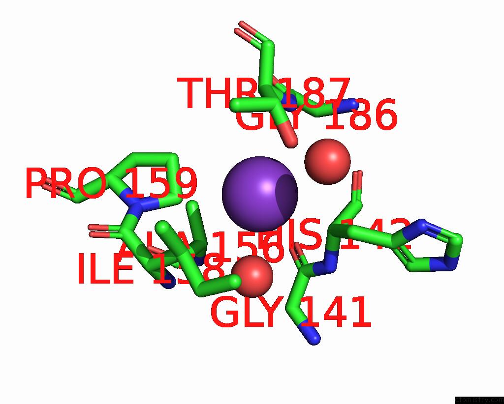
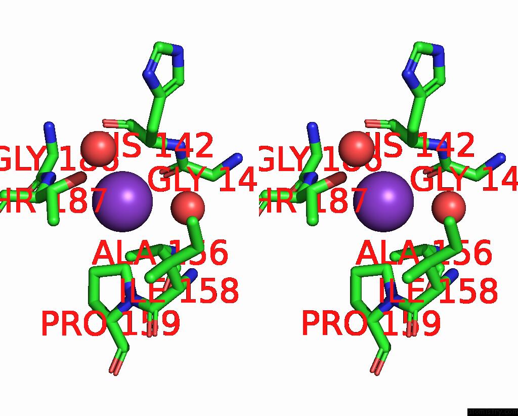
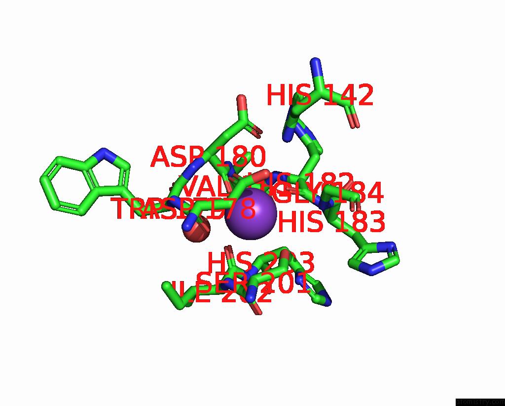
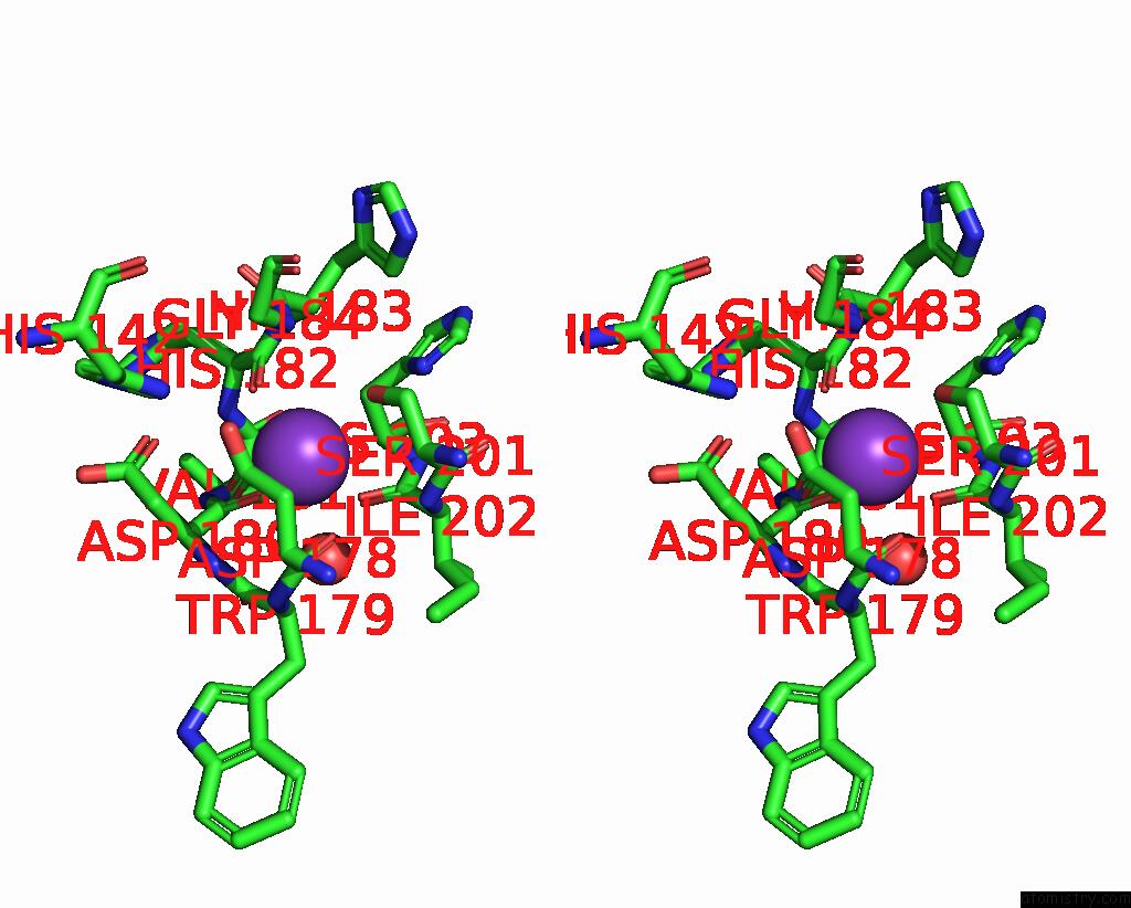
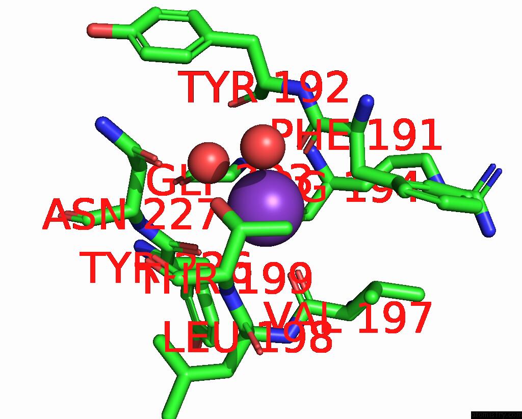
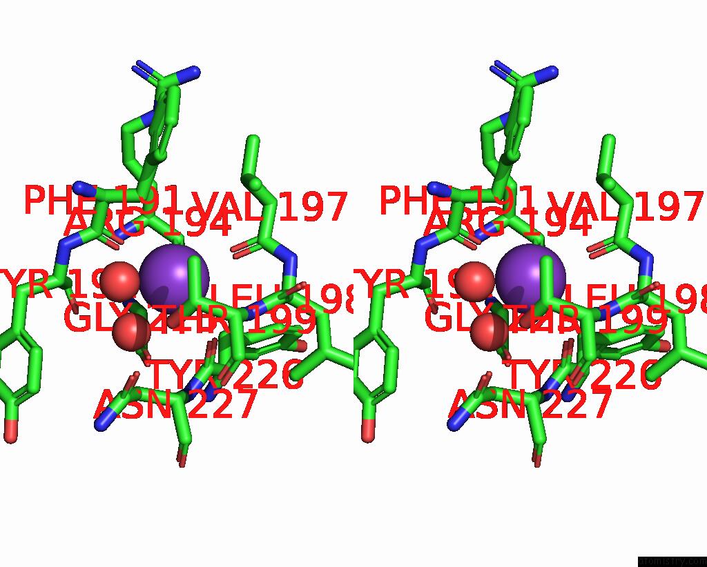
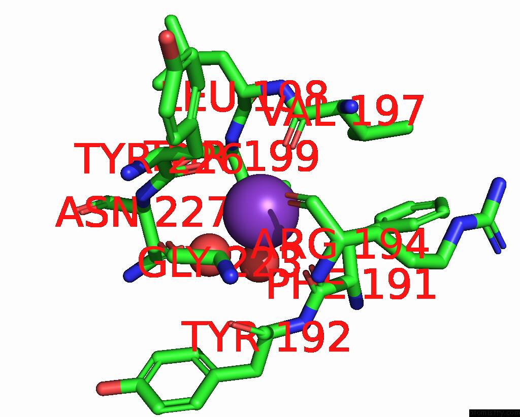
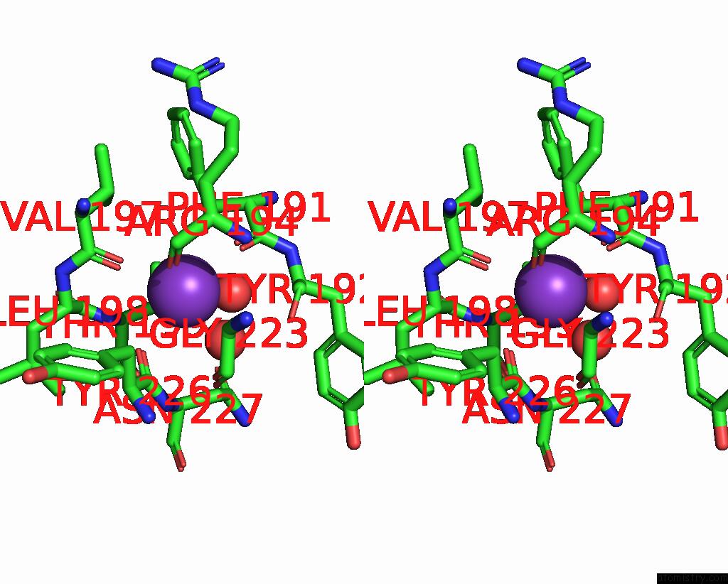
Potassium binding site 1 out of 10 in 9gkw
Go back to
Potassium binding site 1 out
of 10 in the Crystal Structure of Dimethoate Hydrolase (Dmha) of Rhizorhabdus Wittichii in Complex with Octanoic Acid
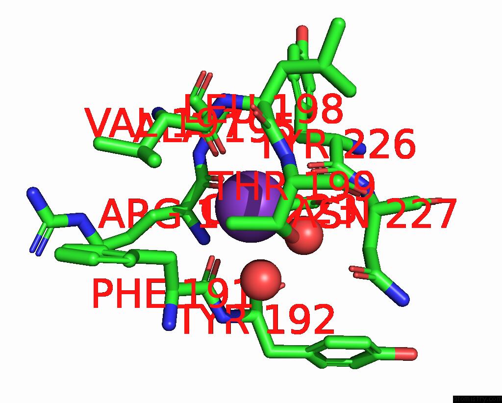
Mono view
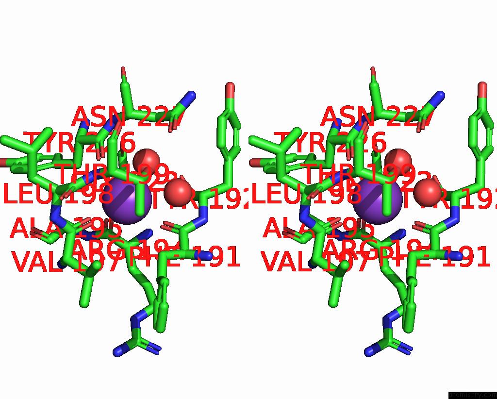
Stereo pair view
Mono view
Stereo pair view
A full contact list of Potassium with other atoms in the K binding
site number 1 of Crystal Structure of Dimethoate Hydrolase (Dmha) of Rhizorhabdus Wittichii in Complex with Octanoic Acid within 5.0Å range:
|
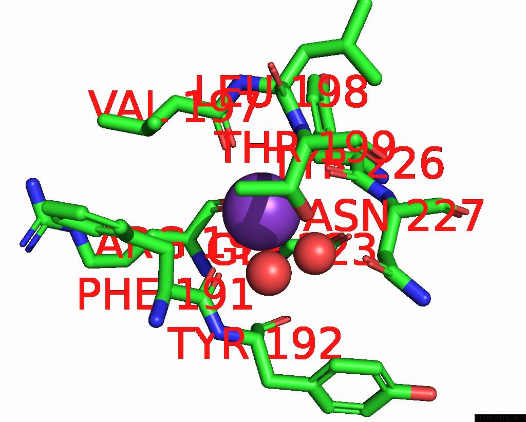
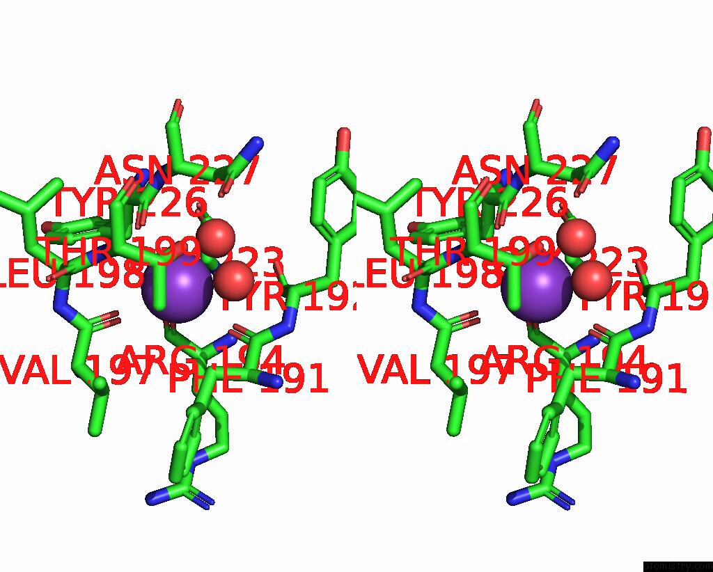
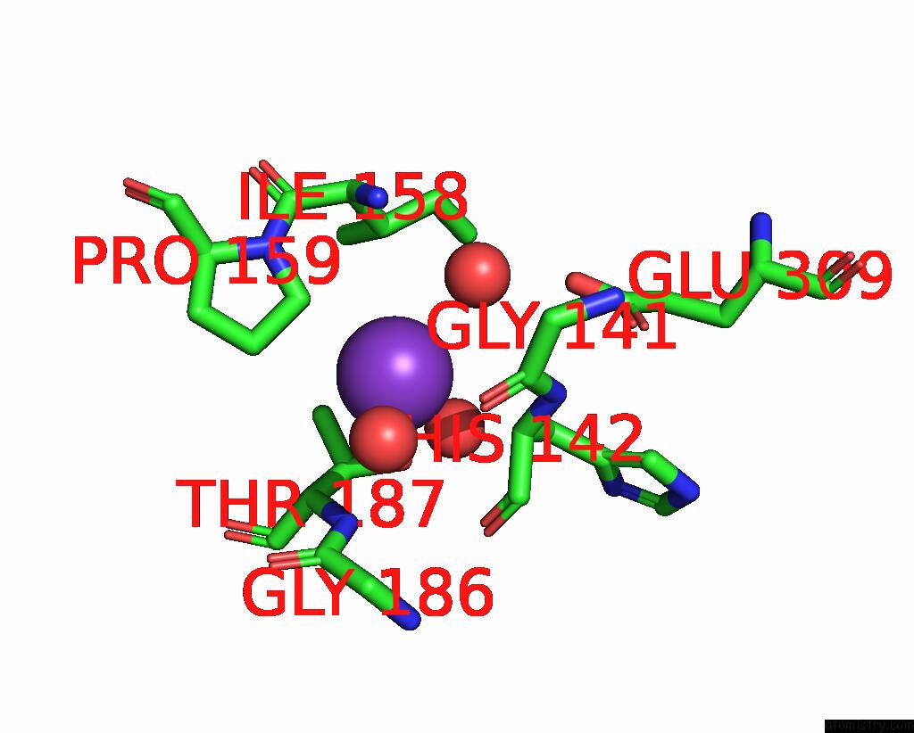
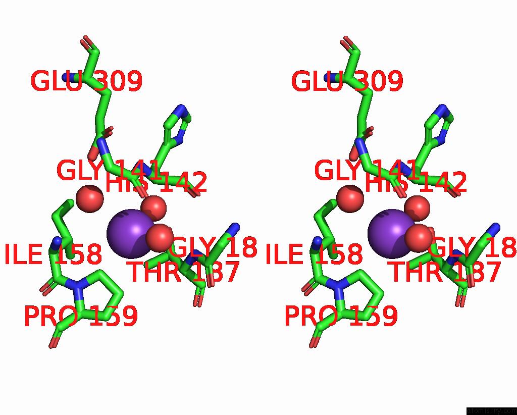
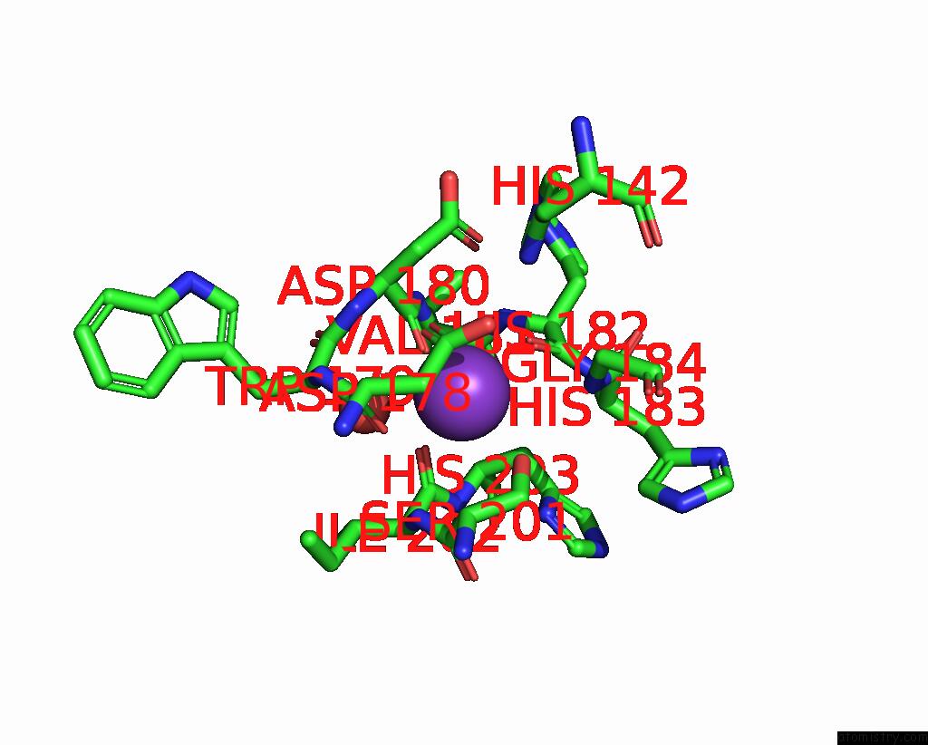
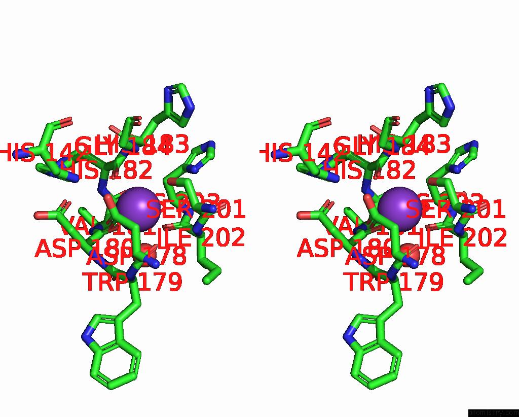
Potassium binding site 2 out of 10 in 9gkw
Go back to
Potassium binding site 2 out
of 10 in the Crystal Structure of Dimethoate Hydrolase (Dmha) of Rhizorhabdus Wittichii in Complex with Octanoic Acid
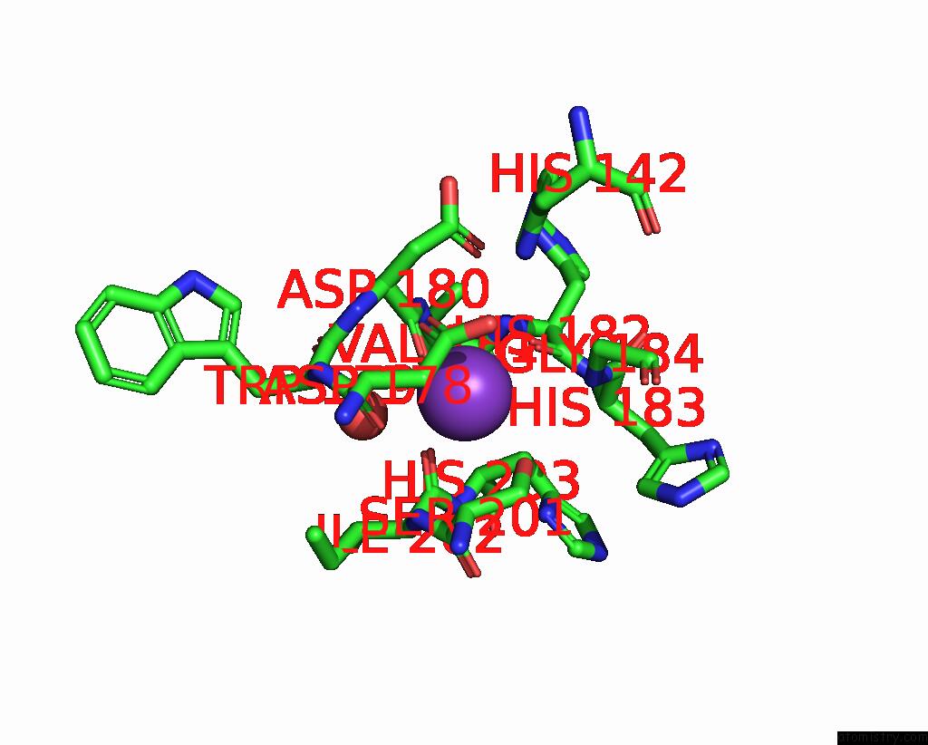
Mono view
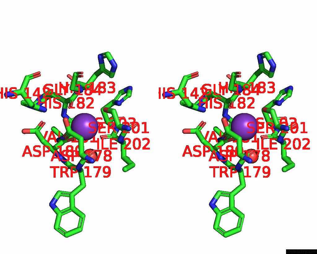
Stereo pair view
Mono view
Stereo pair view
A full contact list of Potassium with other atoms in the K binding
site number 2 of Crystal Structure of Dimethoate Hydrolase (Dmha) of Rhizorhabdus Wittichii in Complex with Octanoic Acid within 5.0Å range:
|
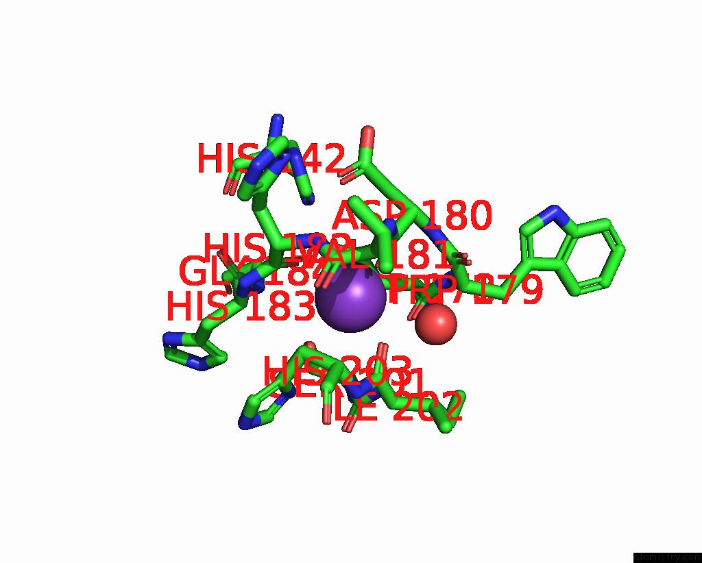
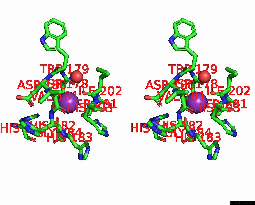
Potassium binding site 3 out of 10 in 9gkw
Go back to
Potassium binding site 3 out
of 10 in the Crystal Structure of Dimethoate Hydrolase (Dmha) of Rhizorhabdus Wittichii in Complex with Octanoic Acid
Mono view
Stereo pair view
Mono view
Stereo pair view
A full contact list of Potassium with other atoms in the K binding
site number 3 of Crystal Structure of Dimethoate Hydrolase (Dmha) of Rhizorhabdus Wittichii in Complex with Octanoic Acid within 5.0Å range:
|
Potassium binding site 4 out of 10 in 9gkw
Go back to
Potassium binding site 4 out
of 10 in the Crystal Structure of Dimethoate Hydrolase (Dmha) of Rhizorhabdus Wittichii in Complex with Octanoic Acid
Mono view
Stereo pair view
Mono view
Stereo pair view
A full contact list of Potassium with other atoms in the K binding
site number 4 of Crystal Structure of Dimethoate Hydrolase (Dmha) of Rhizorhabdus Wittichii in Complex with Octanoic Acid within 5.0Å range:
|
Potassium binding site 5 out of 10 in 9gkw
Go back to
Potassium binding site 5 out
of 10 in the Crystal Structure of Dimethoate Hydrolase (Dmha) of Rhizorhabdus Wittichii in Complex with Octanoic Acid
Mono view
Stereo pair view
Mono view
Stereo pair view
A full contact list of Potassium with other atoms in the K binding
site number 5 of Crystal Structure of Dimethoate Hydrolase (Dmha) of Rhizorhabdus Wittichii in Complex with Octanoic Acid within 5.0Å range:
|
Potassium binding site 6 out of 10 in 9gkw
Go back to
Potassium binding site 6 out
of 10 in the Crystal Structure of Dimethoate Hydrolase (Dmha) of Rhizorhabdus Wittichii in Complex with Octanoic Acid
Mono view
Stereo pair view
Mono view
Stereo pair view
A full contact list of Potassium with other atoms in the K binding
site number 6 of Crystal Structure of Dimethoate Hydrolase (Dmha) of Rhizorhabdus Wittichii in Complex with Octanoic Acid within 5.0Å range:
|
Potassium binding site 7 out of 10 in 9gkw
Go back to
Potassium binding site 7 out
of 10 in the Crystal Structure of Dimethoate Hydrolase (Dmha) of Rhizorhabdus Wittichii in Complex with Octanoic Acid
Mono view
Stereo pair view
Mono view
Stereo pair view
A full contact list of Potassium with other atoms in the K binding
site number 7 of Crystal Structure of Dimethoate Hydrolase (Dmha) of Rhizorhabdus Wittichii in Complex with Octanoic Acid within 5.0Å range:
|
Potassium binding site 8 out of 10 in 9gkw
Go back to
Potassium binding site 8 out
of 10 in the Crystal Structure of Dimethoate Hydrolase (Dmha) of Rhizorhabdus Wittichii in Complex with Octanoic Acid
Mono view
Stereo pair view
Mono view
Stereo pair view
A full contact list of Potassium with other atoms in the K binding
site number 8 of Crystal Structure of Dimethoate Hydrolase (Dmha) of Rhizorhabdus Wittichii in Complex with Octanoic Acid within 5.0Å range:
|
Potassium binding site 9 out of 10 in 9gkw
Go back to
Potassium binding site 9 out
of 10 in the Crystal Structure of Dimethoate Hydrolase (Dmha) of Rhizorhabdus Wittichii in Complex with Octanoic Acid
Mono view
Stereo pair view
Mono view
Stereo pair view
A full contact list of Potassium with other atoms in the K binding
site number 9 of Crystal Structure of Dimethoate Hydrolase (Dmha) of Rhizorhabdus Wittichii in Complex with Octanoic Acid within 5.0Å range:
|
Potassium binding site 10 out of 10 in 9gkw
Go back to
Potassium binding site 10 out
of 10 in the Crystal Structure of Dimethoate Hydrolase (Dmha) of Rhizorhabdus Wittichii in Complex with Octanoic Acid
Mono view
Stereo pair view
Mono view
Stereo pair view
A full contact list of Potassium with other atoms in the K binding
site number 10 of Crystal Structure of Dimethoate Hydrolase (Dmha) of Rhizorhabdus Wittichii in Complex with Octanoic Acid within 5.0Å range:
|
Reference:
L.G.Graf,
C.Moreno-Yruela,
C.Qin,
S.Schulze,
G.J.Palm,
O.Schmoeker,
N.Wang,
D.Hocking,
L.Jebeli,
B.Girbardt,
L.Berndt,
D.M.Weis,
M.Janetzky,
D.Zuehlke,
S.Sievers,
R.A.Strugnell,
C.A.Olsen,
K.Hofmann,
M.Lammers.
Distribution and Diversity of Classical Deacylases in Bacteria Nature Communications 2024.
Page generated: Wed Nov 13 11:17:29 2024
Last articles
Zn in 9IRQZn in 9IYX
Zn in 9J8P
Zn in 9IUU
Zn in 9GBF
Zn in 9G2V
Zn in 9G2L
Zn in 9G2X
Zn in 9G2Z
Zn in 9G2K